BG
BG  BY
BY  CY
CY  CZ
CZ  DE
DE  EE
EE  ES
ES  GR
GR  HU
HU  IS
IS  IT
IT  LT
LT  LV
LV  MY
MY  PL
PL  PT
PT  RO
RO  SK
SK  TR
TR  UA
UA  USA
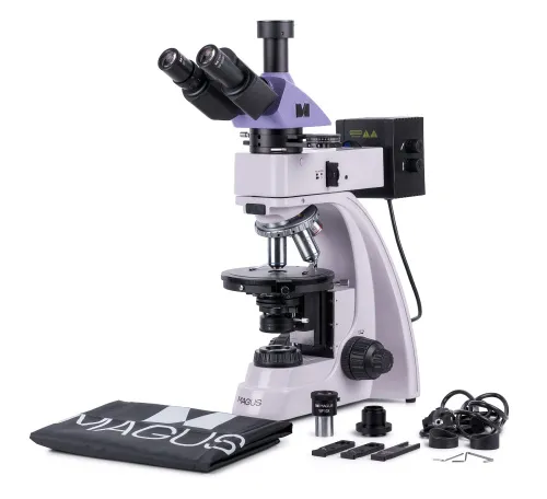
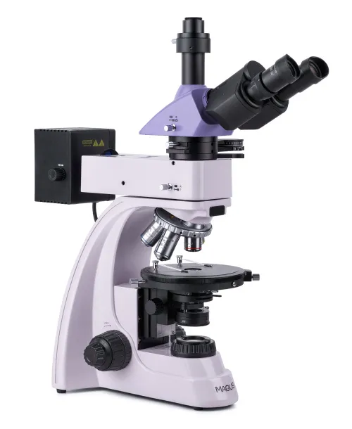
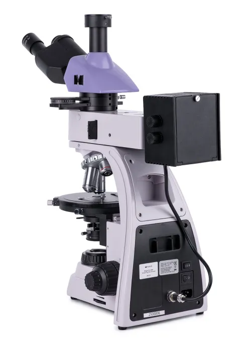
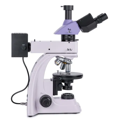
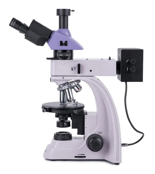
USA MAGUS Pol 850 Polarizing Microscope

Magnification 50–600x. Trinocular head, plan achromatic objectives. Transmitted and reflected light, light sources – 30W halogen bulbs, Köhler illumination in reflected and transmitted light. Bertrand lens and compensators
| Product ID | 82912 |
| Brand | MAGUS |
| Warranty | 5 years |
| EAN | 5905555018539 |
| Package size (LxWxH) | 44x42x67 cm |
| Shipping Weight | 11.5 kg |
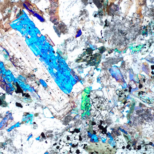
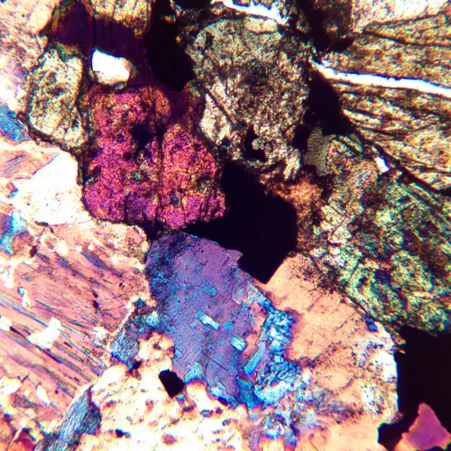
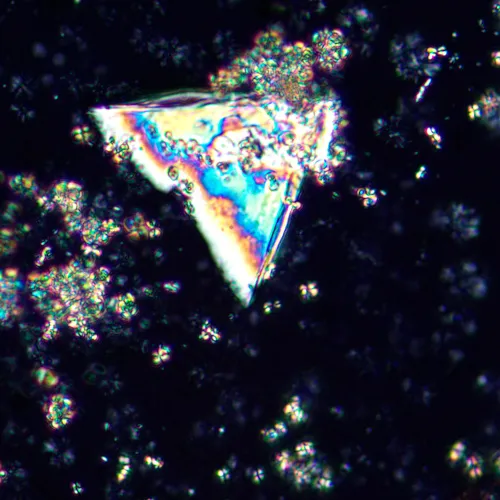
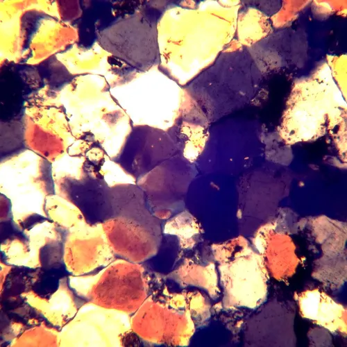
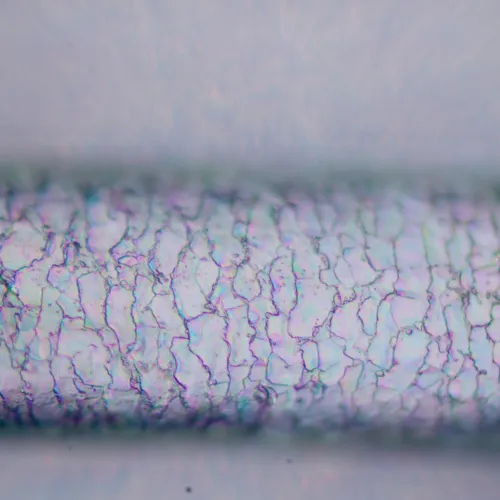
The MAGUS Pol 850 Polarizing Microscope is designed to work in transmitted and reflected light. Available research methods: polarization and brightfield. The microscope is used to study anisotropic biological, geological, and polymer objects as well as opaque polished sections up to 15mm thick with one polished side. The microscope is perfect for a wide range of tasks of professionals in the field of medicine, forensics, mineralogy, crystallography, or petrography.
Optics
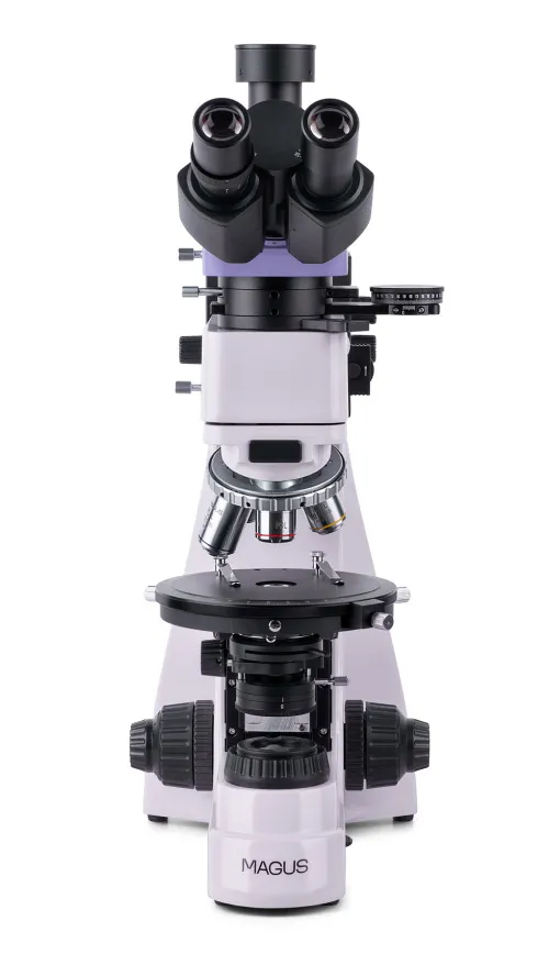
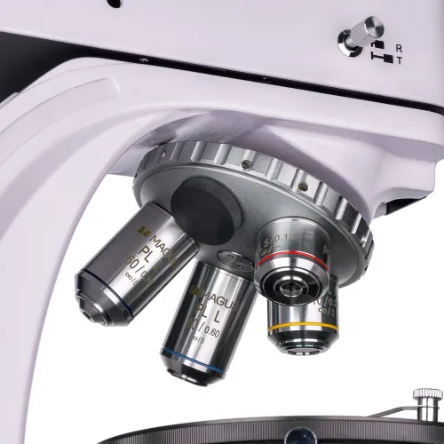
The optics of the microscope are infinity corrected and are free from internal strains. The image formed by the lenses is authentic, free of spurious reflections, high contrast, and detailed. The lenses are installed in a revolving nosepiece with slots centered to the optical axis. There are five slots, with four objectives included in the kit; one additional objective can be installed to obtain further magnification within the magnification range of the microscope. Objectives are designed to work with samples without cover slips.
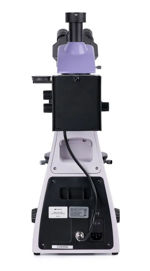
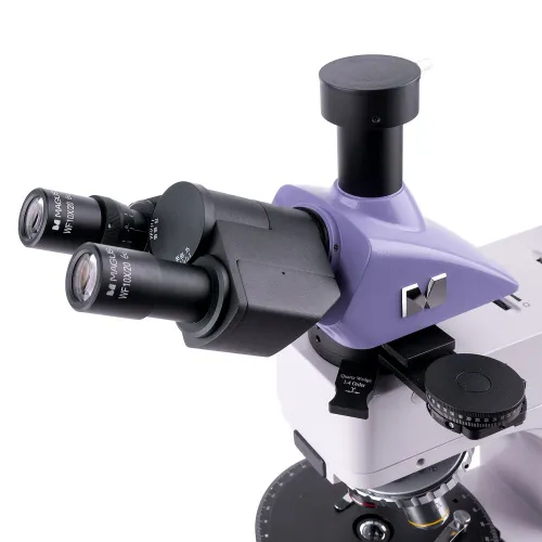
The trinocular head enables you to install a digital camera (not included) in the trinocular tube. The beam splitting is 100:0 or 0:100, i.e., all of the light goes either to the camera or to the eyepiece tubes. The left eyepiece tube has a diopter adjustment ring: It is rotated to compensate for the difference in vision between the eyes and to obtain the clearest possible image of the sample.
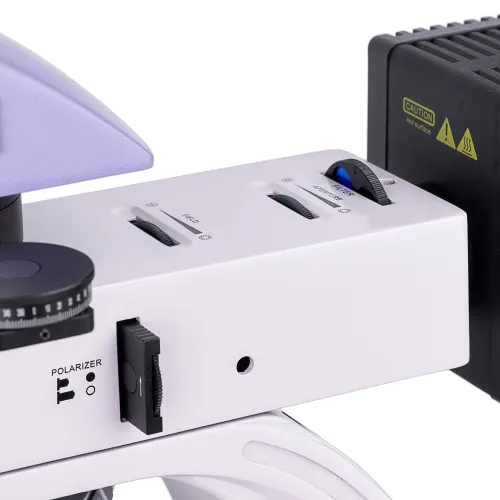
Illumination
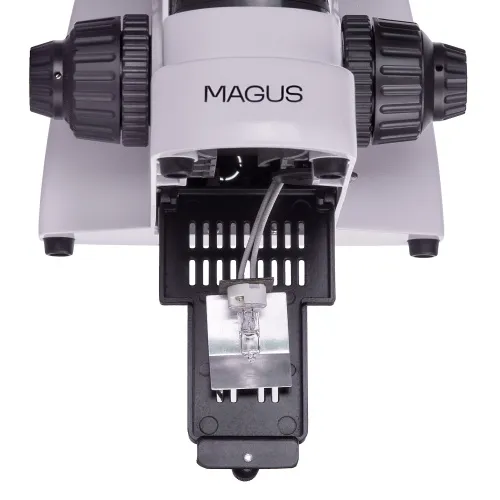
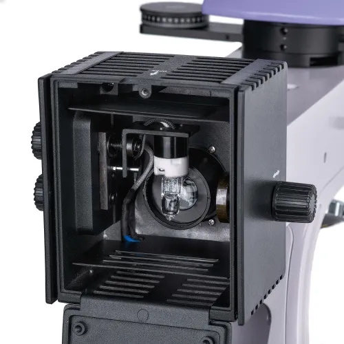
30W halogen bulbs are used for transmitted and reflected light. Their brightness is sufficient to obtain a clearly readable image on each objective, and the “warm” emission spectrum is comfortable during long-term use. Köhler illumination setup will provide increased resolution, and it will remove artifacts in the field of view and darkening at the edges. Light filters are used to accurately adjust color reproduction when working in reflected light.
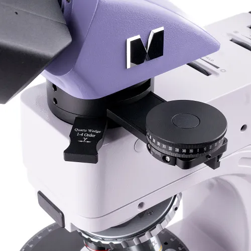
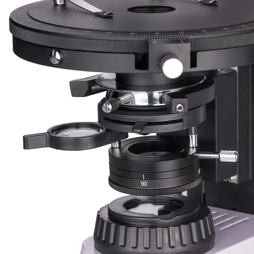
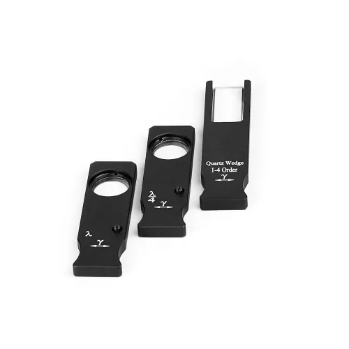
Polarization is available for both transmitted and reflected light. To begin observations in polarized light, an analyzer is introduced into the optical path. Next, the relative position of the analyzer and polarizer is changed by rotating them, which also changes the polarization angle. The contrast of objects is increased with the help of compensators, which are installed in a special slot in the intermediate attachment. The same attachment also contains a Bertrand lens that is used for conoscopic examinations.
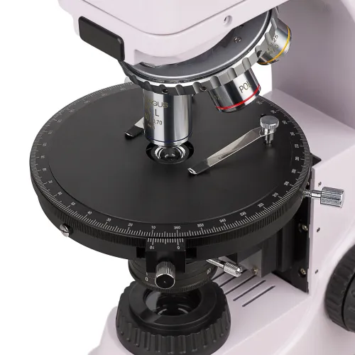
Stage and focusing mechanism
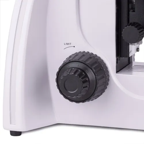
The microscope has a rotatable stage: Turning it changes the way the sample refracts light in polarized light. The stage has a scale on which you can determine the angle of rotation with an accuracy of up to 0.1°. To obtain the brightest and clearest image, the stage can be centered relative to the optical axis of the microscope.
The coarse and fine focusing knobs are located at the bottom of the stand, and so the user may take a relaxed pose with their hands on the table. The coarse tension can be adjusted, and there is also a coarse focusing lock. The fine focusing provides for the smooth movement of the sample.
Accessories
To get the most out of your microscope, use accessories from the MAGUS line: eyepieces, objectives, calibration slides, digital cameras, etc.
Key features:
- For work in transmitted and reflected polarized and natural light
- Trinocular head with a tube for a digital camera; 100:0 or 0:100 beam splitting
- Strain-free plan achromatic objectives for clear images free of artifacts and false internal reflections
- Halogen illumination of the work area, a color spectrum that is comfortable for the eyes; 30W bulb power
- Rotating stage, polarizer, and analyzer with rotation angle marks, Bertrand lens, and compensators
- The revolver slots and stage are centered relative to the optical axis of the microscope
- Compatible with additional accessories
The kit includes:
- Base with a power input, transmitted light source and condenser, focusing mechanism, stage, and revolving nosepiece
- Reflected light illuminator with a lamphouse
- Trinocular head
- Intermediate tube with Bertrand lens, an analyzer and compensator slot
- Compensators: λ compensator, λ/4 compensator, quartz wedge
- Infinity plan achromatic objective: PL 5x/0.12 WD 26.1mm
- Infinity plan achromatic objective: PL 10x/0.25 WD 5.0mm
- Infinity plan achromatic objective: PL 40x/0.6 (spring-loaded) WD 3.98mm
- Infinity plan achromatic objective: PL 60x/0.7 (spring-loaded) WD 2.03mm
- Eyepiece 10x/20 mm (2 pcs.)
- 10x/20mm eyepiece with a scale. Scale value: 0.1mm
- C-mount adapter 1x
- Hex key wrench
- Power cord
- Dust cover
- User manual and warranty card
Available on request:
- 16x/11mm eyepiece (2 pcs.)
- 20x/11mm eyepiece (2 pcs.)
- Infinity plan achromatic objective: PL 2.5x/0.07 WD 11mm
- Infinity plan achromatic objective: PL 50x/0.7 (spring-loaded) WD 3.67mm
- Infinity plan achromatic objective: PL 80x/0.8 (spring-loaded) WD 1.25mm
- Infinity plan achromatic objective: PL 100x/0.85 (spring-loaded) WD 0.4mm
- Mechanical stage attachment
- Digital camera
- Calibration slide
| Product ID | 82912 |
| Brand | MAGUS |
| Warranty | 5 years |
| EAN | 5905555018539 |
| Package size (LxWxH) | 44x42x67 cm |
| Shipping Weight | 11.5 kg |
| Type | biological, light/optical |
| Microscope head type | trinocular |
| Head | Siedentopf |
| Head inclination angle | 30 ° |
| Magnification, x | 50 — 600 |
| Magnification, x (optional) | 25–1000/1600/2000 |
| Eyepiece tube diameter, mm | 23.2 |
| Eyepieces | 10х/20; 10x/20 with a scale (*optional: 16x/11; 20х/11) |
| Objectives | infinity plan achromatic, strain-free: PL 5x/0.12; PL 10x/0.25; PL 40x/0.6; PL 60x/0.7 (*optional: PL 2.5x/0.07; PL 50x/0.7; PL 80x/0.8; PL 100x/0.85); parfocal height 45mm, designed for studying specimens without cover slips |
| Revolving nosepiece | for 5 objectives, centering |
| Working distance, mm | 26.1 (5x); 5.0 (10x); 3.98 (40x); 2.03 (60x); 11 (2.5x); 3.67 (50x); 1.25 (80x); 0.4 (100x) |
| Stage, mm | Ø150 |
| Stage features | 360 ° rotatable, centering, 1° graduation of the rotation angle; a vernier scale for measuring angles with an accuracy of 0.1° |
| Eyepiece diopter adjustment, diopters | ±5 (on the left tube) |
| Condenser | Abbe condenser NA 1.25 with an adjustable aperture diaphragm and a swivel lens |
| Diaphragm | adjustable aperture diaphragm, adjustable iris field diaphragm |
| Focus | coaxial, coarse focusing (21mm, 39.8mm/circle, with a lock knob and tension adjusting knob) and fine focusing (0.002mm) |
| Illumination | halogen |
| Brightness adjustment | ✓ |
| Power supply | 220±22V, 50Hz, AC network |
| Light source type | reflected and transmitted light: 12V/30W |
| Light filters | reflected light: matt, yellow, green, blue |
| Operating temperature range, °C | 5...+35 |
| User level | experienced users, professionals |
| Assembly and installation difficulty level | complicated |
| Polarizer | transmitted light: with 0°, 90°, 180°, 270° marks on the scale; 360° rotatable; reflected light: removable |
| Intermediate tube | built-in analyzer with rotation 0–360°; a vernier scale for measuring angles with an accuracy of 0.1°; Bertrand lens; slot for installing compensators |
| Compensator | λ compensator; λ/4 compensator; quartz wedge |
| Application | laboratory/medical |
| Illumination location | dual |
| Research method | bright field, polarization |
| Pouch/case/bag in set | dust cover |
We have gathered answers to the most frequently asked questions to help you sort things out
Find out why studying eyes under a microscope is entertaining; how insects’ and arachnids’ eyes differ and what the best way is to observe such an interesting specimen
Read this review to learn how to observe human hair, what different hair looks like under a microscope and what magnification is required for observations
Learn what a numerical aperture is and how to choose a suitable objective lens for your microscope here
Learn what a spider looks like under microscope, when the best time is to take photos of it, how to study it properly at magnification and more interesting facts about observing insects and arachnids
This review for beginner explorers of the micro world introduces you to the optical, illuminating and mechanical parts of a microscope and their functions
Short article about Paramecium caudatum - a microorganism that is interesting to observe through any microscope