All novices tend to have some difficulty when starting work with a microscope. So if you have recently bought a microscope, it is very likely that you have some reasonable and perfectly understandable questions. What magnification do I need? Does the field of view play an important role? Why do I need additional eyepieces? We have gathered answers to the most frequently asked questions to help you sort things out. This information will be helpful not only for people who already own a microscope but also for those who are just planning to buy this optical instrument.
“Why is it important to start observation with 4x magnification?”
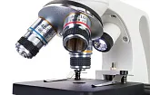
A typical compound optical microscope has three or four objective lenses with 4x, 10x, 40x and 100x (oil immersion) magnification. 4x objective provides the smallest increase, so you will be able to observe a large area of the sample. This allows you to easily find the desired area for further microscopic observation. When you have found the area, place it in the center of the field of view and switch to a higher magnification objective. It is much easier to focus the view using a low-power objective lens, rather than a powerful one
Back to top
“How to use a microscope as a measuring tool?”
You can measure the size of a sample using a microscope, but first it is necessary to calibrate the microscope. This requires a micrometer scale (called a reticule) integrated in the microscope eyepiece and a measuring scale on the stage. The reticule has divisions, but you can’t use them to measure the samples directly. Let’s say a stage scale has a scale value equal to 0.01 mm. If you line up the scale on the stage with the eyepiece scale, you will see that X divisions on the eyepiece scale correspond to Y divisions of the measuring scale on the stage. To calculate the division value of the eyepiece scale, the following formula is used:
Eyepiece division value = 0.01 * Y/X
It is necessary to perform calculations like these for each possible combination of eyepieces and objective lenses as they provide different magnifications.
Back to top
“How to calculate the magnification of a microscope?”
To calculate the magnification of a microscope, simply multiply the magnification of the microscope eyepiece by the magnification of the objective lens. The total magnification of a typical compound microscope with 10x eyepiece and 4x, 10x, 40x, 100x objectives will be 40x, 100x, 400x and 1000x depending on the lenses used. The same principle applies to stereo microscopes. Some stereo microscopes are equipped with objectives that have a variable magnification in the range of 0.75x - 7.5x. The total magnification of the microscope with a 10x eyepiece will vary accordingly from 7.5 to 75-fold; with a 25x eyepiece - from 18.75 to 187.5-fold.
Back to top
“How does magnification of a microscope relate to the focal length?”
In microscopy, the focal length is understood as a distance between the objective lens and the upper part of the observed object. The focal length of the optical system shows how efficiently the system collects and focuses light rays. Commonly, the greater the magnification of the microscope, the shorter its focal length.
Back to top
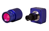
“How to take photos through a microscope?”
There are several different ways to take photos with a microscope. Many modern microscopes are equipped with built-in digital cameras, which are connected to a PC via USB-cable. It’s very convenient to take photos with such microscope models. Even if your microscope does not have a built-in digital camera, the possibility to make photos still remains. There is a large number of microscope camera models that can be installed in place of an eyepiece. Using a special adapter, you can also attach a compact camera or even DSLR camera to your microscope. Compact cameras are mounted so that their objective lens "looks" into the microscope eyepiece. DSLR cameras work by replacing the camera’s objective lens with an eyepiece tube adapter.
Back to top
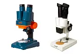
“What is the difference between low-powered and high-powered microscopes?”
This question is a little tricky since the definitions are slightly blurred. For example, optical (light) microscopes are usually equipped with four objectives: 4x and 10x are low power objectives; 40x and 100х are powerful ones. The total magnification (received with 10x eyepiece) of less than 400x characterizes the microscope as a low-powered model; more than 400x as a powerful one. As a typical compound microscope gives a greater increase than a stereo microscope (or dissecting microscope), some sources refer to stereo models as part of the low-powered microscope group, and compound microscopes as powerful models.
Back to top
“We are looking for a teaching microscope. Is it better to choose a model with three or four objectives?”
It depends on the purpose of your observation. Basic educational microscopes usually come with three objective lenses (4x, 10x and 40x). The maximum magnification power of these models is 400x. The advanced teaching microscope models are usually equipped with the fourth objective – 100x oil immersion objective lens. Using this objective with a 10x eyepiece and oil immersion technique you can obtain 1000x increase. 1000x magnification power allows you to observe the target object in great detail. However, such a large increase can cause some difficulties when focusing on objects. The oil immersion technique is required (which can be a laborious task), and, besides, the objective itself is a quite expensive accessory. At 400x magnification you won’t get as detailed images as under 1000x increase, but it is much easier to focus on objects, there is no need to use immersion oil, and you will also save more money.
Back to top
“What is the diopter adjustment?”
The diopter adjustment allows you to focus one eyepiece independently from the other to compensate for the difference in vision between your two eyes. When the diopter adjustment is made correctly both eyes feel comfortable during observations.
Back to top
“What is the field of view in a microscope?”
The field of view is the diameter of the illuminated circle seen through the eyepiece. The higher the magnification, the smaller the field of view. To obtain the exact value of your microscope’s field of view, place a transparent ruler under the microscope objective and count how many millimeter-lines fit inside the visible illuminated circle.
Back to top
“What is the difference between Achromatic, Plan Achromatic, Semi-Apochromatic, and Apochromatic objectives?”
The main difference between the microscope objectives is design, image quality, and research methods. Achromatic objectives feature reduced spherical and chromatic aberrations. This objective type has a flat field of focus in the middle 65% of the field of view. The delivered image may have a blue-red hint. Plan achromatic objectives correct for color aberrations. They make the field of view flat in order to provide a very sharp image across the entire picture. These objective lenses are excellent for macro photography. Semi-apochromatic objectives feature average image quality (in comparison to achromatic and plan achromatic objectives). The optical elements in these objectives contain fluorite that allows for using them in fluorescence microscopy. Apochromatic objectives (in comparison to achromatic objectives) deliver a sharper image in clearer colors. This is achieved through an enlarged spectral region.
Back to top
“Can I see germs with a microscope?”
You won’t be able to observe germs using an amateur microscope. The reason that it’s impossible is because they are too small. You can observe germs only with a microscope that is able to provide at least 1200x magnification power and the specimen has to be stained. So you will need to use a quite powerful microscope model and special methods of sample preparation. Of course, there are giants even among bacteria, which will be visible even at 900x magnification, but they live in the ocean's deepest depths and it's virtually impossible to get these specimens.
Back to top

 BG
BG  BY
BY  CY
CY  CZ
CZ  DE
DE  EE
EE  ES
ES  GR
GR  HU
HU  IS
IS  IT
IT  LT
LT  LV
LV  MY
MY  PL
PL  PT
PT  RO
RO  SK
SK  TR
TR  UA
UA  USA
USA