BG
BG  BY
BY  CY
CY  CZ
CZ  DE
DE  EE
EE  ES
ES  GR
GR  HU
HU  IS
IS  IT
IT  LT
LT  LV
LV  MY
MY  PL
PL  PT
PT  RO
RO  SK
SK  TR
TR  UA
UA  USA
USA MAGUS Lum 400L Fluorescence Microscope
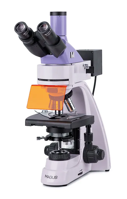
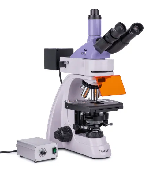
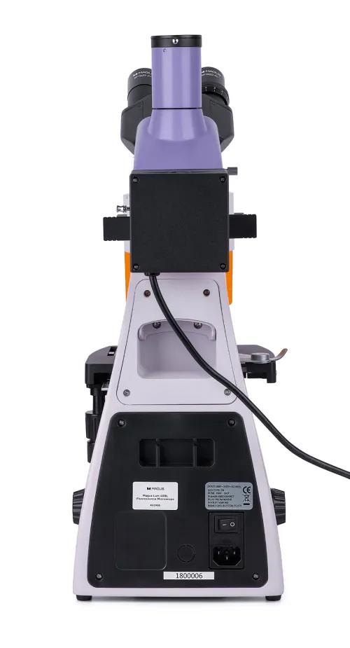
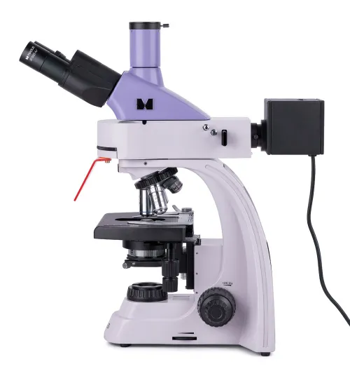
Magnification: 40–1000x. Trinocular head, plan achromatic and plan fluorite objectives, four 5W LEDs of different wavelengths, transmitted light source – 3W LED, Köhler illumination in reflected and transmitted light
| Product ID | 82905 |
| Brand | MAGUS |
| Warranty | 5 years |
| EAN | 5905555018171 |
| Package size (LxWxH) | 45x30x96 cm |
| Shipping Weight | 14.5 kg |
The MAGUS Lum 400L Fluorescence Microscope can be used for two microscopy techniques: fluorescence in reflected light and brightfield in transmitted light. When equipped with additional accessories, you can use the microscope to conduct research in polarized light as well as use darkfield and phase contrast techniques. The microscope can be used for research and laboratory diagnostics in the fields of medicine, pharmacology, forensics, biotechnology, veterinary medicine, etc.
Optics
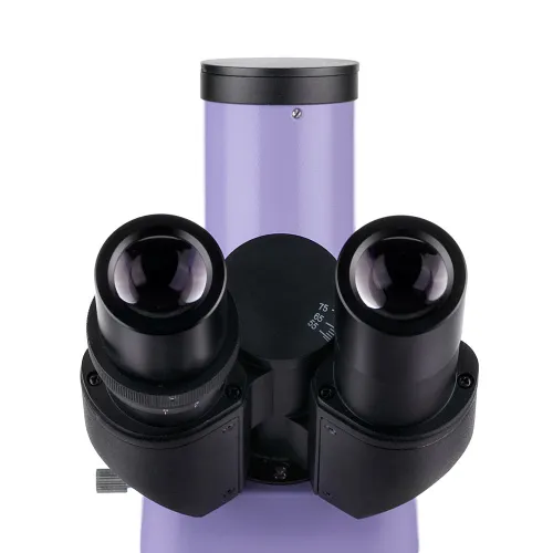
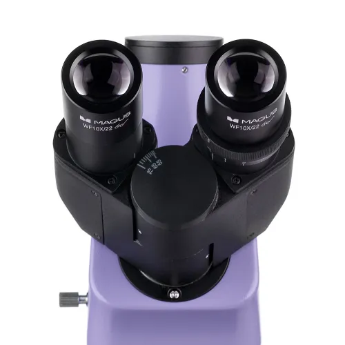
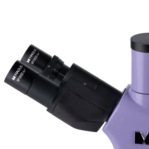
The trinocular head includes two eyepiece tubes for observations and a trinocular tube for mounting a digital camera (not included). For comfortable longer observations, the eyepiece tubes can be adjusted to match the height of the observer, as they rotate 360°. They are 30° inclined.
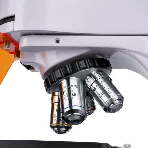
The 5 objectives revolving nosepiece is oriented toward the interior – the user can see the objective inserted into the optical path. It comes with four infinity plan achromatic objectives: three for brightfield and one for fluorescence technique (fluo). One slot is left free when all objectives are installed in the revolving nosepiece. It is used for adjusting the position of the reflected light illuminator. You can also install an optional objective to get extra magnification.
Illumination
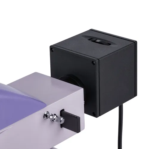
The reflected light – four 5W LEDs. Each LED has in a specific waveband: blue (B), green (G), violet (V), and ultraviolet (UV). To choose the working LED, you have to rotate the ring. The advantage of LED illumination is that LEDs get bright quickly and turn off quickly, making it easy to switch from brightfield to fluorescence and back again. LEDs do not heat up, they do not need a heat sink, and they have a lifetime of 50,000 hours without replacement. LED-based fluorescence is an easy to maintain and safe microscopy technique and, therefore, it is recommended for use at schools and universities.
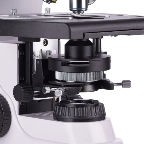
The transmitted light is produced by a 3W LED. It also has a lifespan of 50,000 hours. The illumination system also includes a condenser. The condenser has a slot for a darkfield slider or a phase contrast slider. A slider is an easy and quick way to change the microscopy technique without changing the condenser.
For both transmitted and reflected light, you can set up Köhler illumination. The object of study is in sharp focus, and the image artifacts are removed. There is no darkening at the edges and the resolution limit can be reached at each objective.
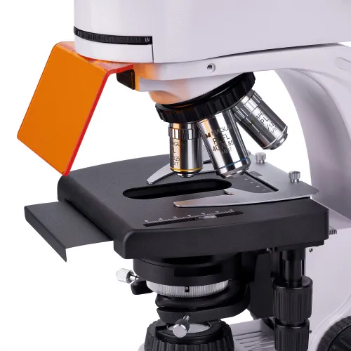
Stage and focusing mechanism
The microscope stage is equipped with a removable specimen holder, which can be removed for manual scanning. The table does not have a positioning rack. Therefore, it requires less space for work. A black stage plate can be installed on the stage to eliminate stray light from the transmitted light source when observing in reflected light.
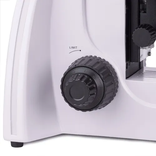
Coaxial coarse and fine focusing knobs are located at the base of the microscope on both sides. Coarse focusing has a lock knob and tension adjustment.
Accessories
The MAGUS Lum 400L microscope has a wide range of compatible accessories that can be used to expand its capabilities. It includes eyepieces, objectives, darkfield, polarized light and phase contrast devices as well as digital cameras and calibration slides.
Key features:
- Study of samples in reflected light using the fluorescence microscopy technique and in transmitted light using the brightfield microscopy technique.
- Infinity plan achromatic objectives: three brightfield objectives, one fluo objective
- Trinocular head, 360° rotatable barrels; a trinocular tube for mounting a digital camera
- Four reflected light LEDs operating at different wavelengths: blue (B), green (G), violet (V) and ultraviolet (UV)
- Transmitted light 3W LED, Köhler transmitted and reflected light illumination
- Stage without a positioning rack, removable specimen holder
- Wide range of accessories, including darkfield, polarized light and phase contrast devices
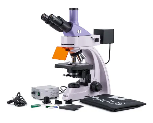
The kit includes:
- Stand with built-in power supply, transmitted light source, focusing mechanism, stage, condenser mount, and revolving nosepiece
- Abbe condenser
- Reflected light illuminator
- Lamphouse with LEDs
- Trinocular head
- Infinity plan achromatic objective: PL 4x/0.10 WD 19.8mm
- Infinity plan achromatic objective: PL 10x/0.25 WD 5.0mm
- Infinity plan achromatic objective, fluo: PL FL 40x/0.85 (spring-loaded) WD 0.42mm
- Infinity plan achromatic objective: PL 100x/1.25 (spring-loaded, oil) WD 0.36mm
- Eyepiece 10x/22mm with long eye relief (2 pcs.)
- Eyepiece eyecup (2 pcs.)
- UV shield
- Filter slider
- Black protective plate for under-stage installation
- C-mount camera adapter
- Color filter (4 pcs.)
- Bottle of immersion oil
- Reflected light illuminator power supply
- Power cord
- Reflected light illuminator power cord
- Power cord of the reflected light illuminator power supply
- Dust cover
- User manual and warranty card
Available on request:
- 10x/22mm eyepiece with a scale
- 12.5x/14mm eyepiece (2 pcs.)
- 15x/15mm eyepiece (2 pcs.)
- 20x/12mm eyepiece (2 pcs.)
- 25x/9mm eyepiece (2 pcs.)
- Infinity plan achromatic objective, fluo: PL FL 10x/0.35 WD 2.37mm
- Infinity plan achromatic objective: PL 20х/0.40 WD 8.8mm
- Infinity plan achromatic objective: PL 60x/0.80 ∞/0.17 WD 0.46mm
- Phase contrast device
- Darkfield condenser NA 0.9
- Oil darkfield condenser NA 1.36–1.25Darkfield slider
- Polarization device
- Digital camera
- Calibration slide
- LCD Monitor
| Product ID | 82905 |
| Brand | MAGUS |
| Warranty | 5 years |
| EAN | 5905555018171 |
| Package size (LxWxH) | 45x30x96 cm |
| Shipping Weight | 14.5 kg |
| Type | biological, light/optical |
| Microscope head type | trinocular |
| Head | Gemel head (Siedentopf, 360° rotation) |
| Head inclination angle | 30 ° |
| Magnification, x | 40 — 1000 |
| Magnification, x (optional) | 40–1250/1500/2000/2500 |
| Eyepiece tube diameter, mm | 30 |
| Eyepieces | 10х/22mm, eye relief: 10mm (*optional: 10x/22mm with scale, 12.5x/14; 15x/15; 20x/12; 25x/9) |
| Objectives | infinity plan achromatic and fluo objectives: PL 4x/0.10, PL 10x/0.25, PL FL 40x/0.85, PL 100x/1.25 (oil); parfocal distance 45mm (*optional: PL FL 10x/0.35, PL 20х/0.40, PL 60x/0.80 ∞/0.17) |
| Revolving nosepiece | for 5 objectives |
| Working distance, mm | 19.8 (4x); 5.0 (10x); 0.42 (FL 40x); 0.36 (100x); 2.37 (FL 10х); 8.8 (20х); 0.46 (60х) |
| Interpupillary distance, mm | 48 — 75 |
| Stage, mm | 180x150 |
| Stage moving range, mm | 75/50 |
| Stage features | two-axis mechanical stage, without a positioning rack |
| Condenser | Abbe condenser, N.A. 1.25, center-adjustable, height-adjustable, adjustable aperture diaphragm, a slot for a darkfield slider and phase contrast slider, dovetail mount |
| Diaphragm | adjustable aperture diaphragm, adjustable iris field diaphragm |
| Focus | coaxial, coarse focusing (21mm, 39.8mm/circle, with a lock knob and tension adjusting knob) and fine focusing (0.002mm) |
| Illumination | LED, fluorescent |
| Brightness adjustment | ✓ |
| Power supply | 85–265V, 50/60Hz, AC network |
| Light source type | reflected light: 4 LEDs of different wavelengths, 5W; transmitted light: 3W LED |
| Light filters | yes |
| Ability to connect additional equipment | darkfield slider, phase contrast device (condenser and objectives), darkfield condenser (dry or oil), polarization devices (polarizer and analyzer) |
| User level | experienced users, professionals |
| Assembly and installation difficulty level | complicated |
| Fluorescent module | filters: ultraviolet (UV), violet (V), blue (B), green (G) |
| Fluorescence filter: filter type, excitation wavelength/dichroic mirror/emission wavelength | ultraviolet (UV), 320–380nm/420nm/435nm; violet (V), 380–415 nm/460 nm/475nm; blue (B), 410–490nm/505nm/515nm; green (G), 475–550nm/580nm/595nm |
| Application | laboratory/medical |
| Illumination location | dual |
| Research method | bright field, fluorescence |
| Pouch/case/bag in set | dust cover |
We have gathered answers to the most frequently asked questions to help you sort things out
Find out why studying eyes under a microscope is entertaining; how insects’ and arachnids’ eyes differ and what the best way is to observe such an interesting specimen
Read this review to learn how to observe human hair, what different hair looks like under a microscope and what magnification is required for observations
Learn what a numerical aperture is and how to choose a suitable objective lens for your microscope here
Learn what a spider looks like under microscope, when the best time is to take photos of it, how to study it properly at magnification and more interesting facts about observing insects and arachnids
This review for beginner explorers of the micro world introduces you to the optical, illuminating and mechanical parts of a microscope and their functions
Short article about Paramecium caudatum - a microorganism that is interesting to observe through any microscope