
 BG
BG  BY
BY  CY
CY  CZ
CZ  DE
DE  EE
EE  ES
ES  GR
GR  HU
HU  IS
IS  IT
IT  LT
LT  LV
LV  MY
MY  PL
PL  PT
PT  RO
RO  SK
SK  TR
TR  UA
UA  USA
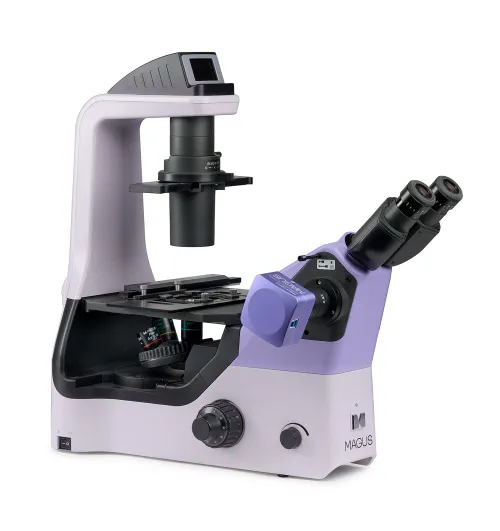
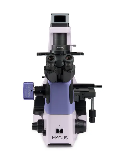
USA MAGUS Bio VD360 Biological Inverted Digital Microscope
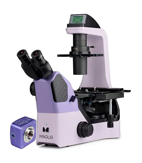
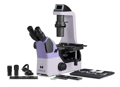
With a camera. Magnification: 40–400x. Binocular microscope head with side camera port, plan achromatic objectives, 3W LED illuminator, condenser with phase slider, emboss contrast device, and intelligent lighting control system
| Product ID | 83278 |
| Brand | MAGUS |
| Warranty | 5 years |
| EAN | 5905555019895 |
| Package size (LxWxH) | 56x65x67 cm |
| Shipping Weight | 25 kg |
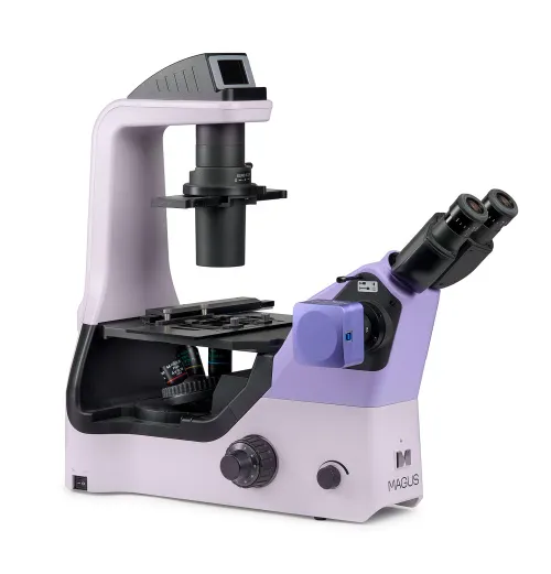
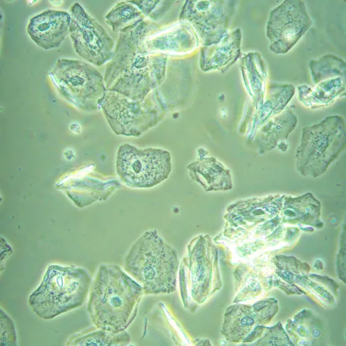
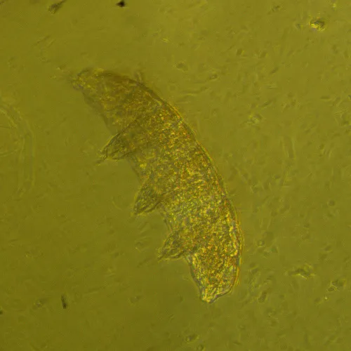
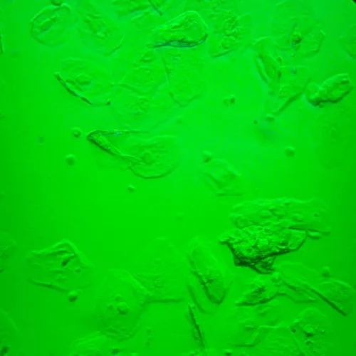
The research-grade microscope. The microscope is designed for studying liquid precipitates, cell colonies, living cells, tissue cultures, and other stained and unstained specimens in laboratory glassware. The inverted design of the microscope involves the use of Petri dishes, multiwell plates, bottles, roller bottles, and flasks up to 75mm with a bottom thickness of 1.2mm. The microscope uses special objectives to work with such glassware. With the condenser removed, you can observe cell cultures in Petri dishes or in cylindrical flasks up to 187mm tall. Observations are made in transmitted light using the brightfield, phase contrast, and emboss contrast techniques. Optional components will allow for the use of the Hoffman modulation contrast techniques. The microscope is equipped with a 5MP camera with a USB 3.0 interface. The camera connects to a computer and displays the image on its screen. The software complements the system with analysis and documentation functions. The camera mounts to the microscope using either the 0.5x or 1x adapter that is included with the microscope. The adapter is selected based on the tasks to be solved. The 0.5x adapter extends the field of view, while the 1x adapter transfers the image “as is”, while cropping the edges, but each detail appears larger on the sensor. The microscope is used for research in medicine, pharmacology, biology, and virology.
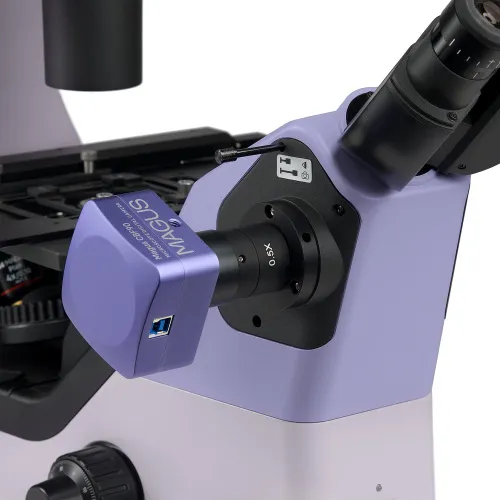
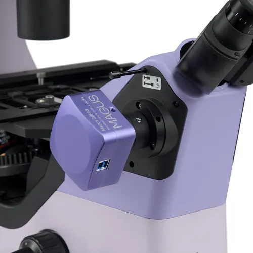
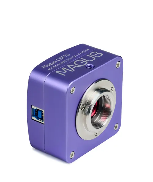
Digital camera
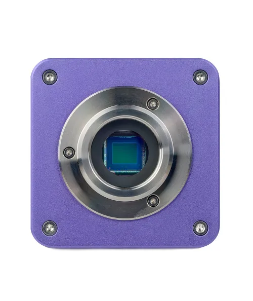
MAGUS CBF90 is a science camera with 5MP resolution and a 2/3" sensor. The camera produces a realistic image in 2448x2048px. Video is recorded at 35fps or 50fps depending on its resolution. Videos are smooth, with soft and subtle transitions between frames. The movement of the sample is displayed in real time with no delays. The camera provides convenient work with moving objects and is suitable for conducting demonstrations in a classroom. The camera is equipped with a USB 3.0 interface. The data transfer rate is 10 times faster than that of USB 2.0 cameras. The high-speed camera is recommended for professional laboratories, research, or university training.
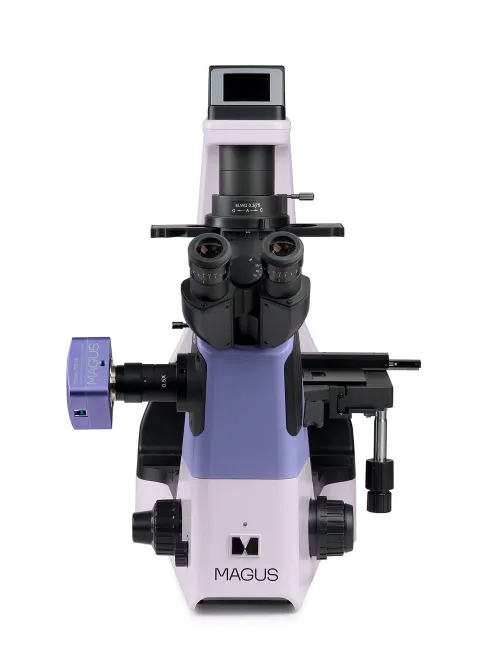
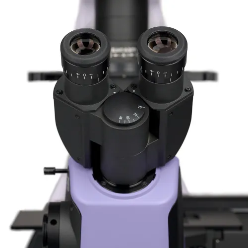
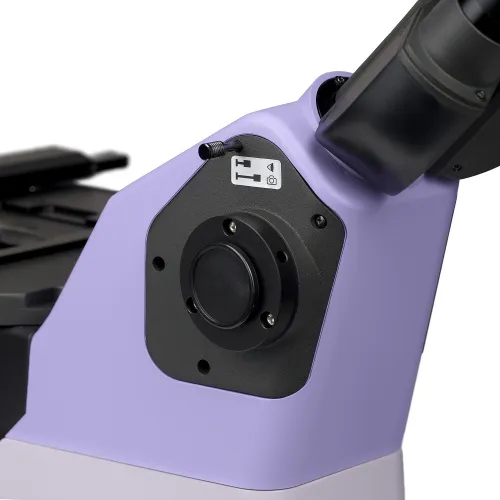
Microscope head
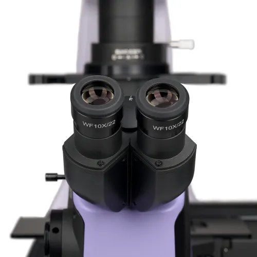
Binocular, with infinity-corrected optics. The digital camera is mounted in the side camera port on the microscope head. The light splitting ratio is 100/0 or 0/100. A slot with a plug is intended for installing the emboss contrast slider.
Revolving nosepiece
Coded, for 5 objectives. An additional objective can also be installed in the free slot in order to achieve extra magnification.
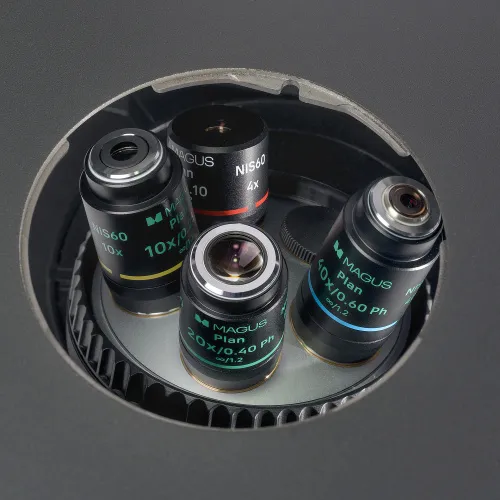
Objectives
Infinity-corrected plan achromatic objectives with an extended working distance have been corrected for a dish bottom thickness of 1.2mm. The parfocal distance is 60mm. The 4x objectives are used for the brightfield technique. The 10х, 20х, and 40х objectives are phase objectives that differ from the 4x objectives by having a phase ring in the plane of the exit pupil. These objectives are designed for phase contrast observations, but they are also suitable for the brightfield and emboss contrast techniques.
Maintaining comfortable brightness levels when switching magnifications
The objectives of different magnifications transmit light with different levels of intensity, and so each time you change objectives, the brightness of the light must be adjusted. Switching from a higher to a lower magnification objective causes eye fatigue, as the image brightness in the eyepieces increases sharply. The MAGUS Bio VD360, which is equipped with intelligent brightness control, solves this problem. The microscope remembers the brightness for each objective that the user has selected and automatically sets this brightness when turning the nosepiece. Intelligent control reduces the time required to adjust brightness. MAGUS Bio VD360 increases user comfort and saves time even when work requires frequent magnification changes.
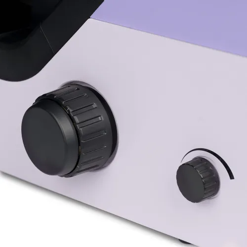
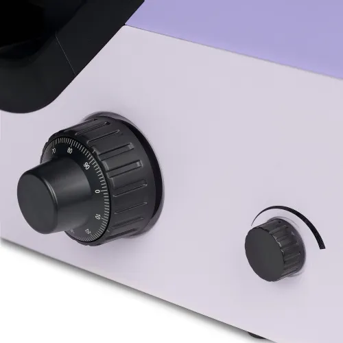
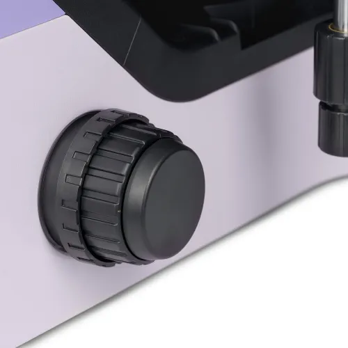
Focusing mechanism
The coarse and fine focusing knobs are coaxial and located low. The researcher can place their hands on the table and take a comfortable position in front of the microscope. The ring on the right side adjusts the tension of the coarse focusing travel. The user adjusts the comfortable tension for work.
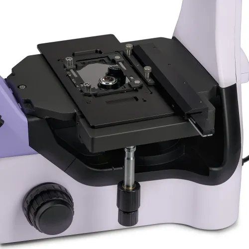
Stage
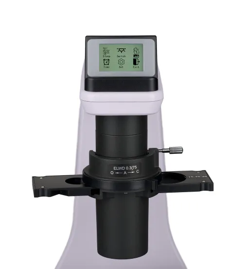
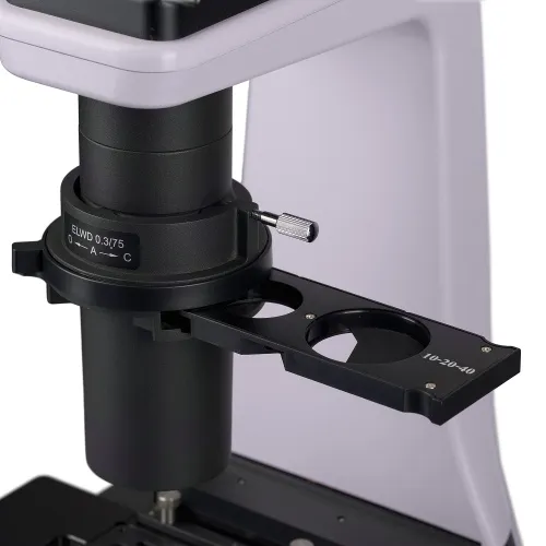
The stage is fixed. A special mechanism is installed on the stage, which moves laboratory glassware in two mutually perpendicular directions. The smooth and subtle movement of the object augments the accuracy of the study: no part of the specimen will be overlooked. The long stage control handle ensures user comfort: The hand rests on the table with no strain. The microscope kit includes a universal labware holder.
Condenser
The condenser has 0.3 NA and a working distance of 75mm. A phase contrast slider or an emboss contrast slider with a sector diaphragm is installed in the condenser slot. A phase contrast slider is a plate with three holes. One hole has a phase insert for 10x, 20x, and 40x objectives, while the other two are used for brightfield observations. The annular diaphragm of the slider is aligned with the phase ring of the objective using a centering telescope. An emboss contrast slider with a sector diaphragm is also a plate with three holes. The emboss contrast microscopy is enabled by introducing the sector diaphragm into the optical path and setting the slider in the microscope head slot to the position that matches the objective being used. The green filter (ND6), when placed into the slot with the sector diaphragm, removes color halos at the edges of the observed structures. The slider’s free holes are designed for brightfield observations: one for a 4x objective and the second one for 10х, 20х, and 40х objectives.
Light source
The 3W LED delivers bright specimen illumination that is sufficient for all of the microscope’s available techniques and objectives. The color temperature does not change when you adjust the brightness. The LED lifetime is 50,000 hours.
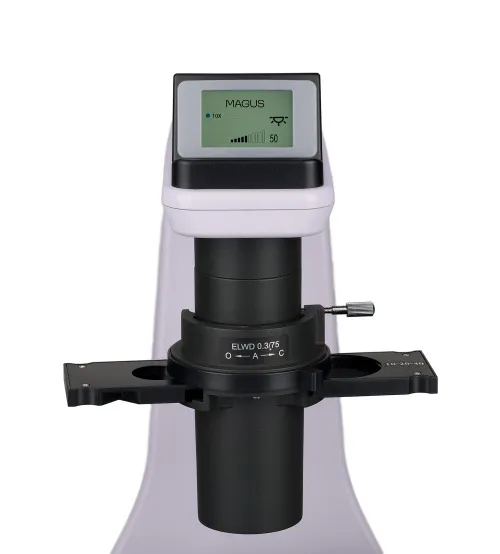
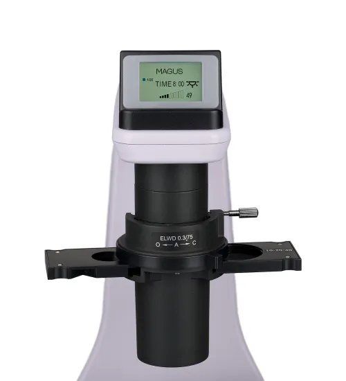
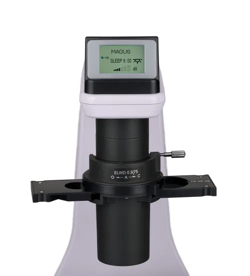
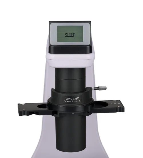
Status LCD screen
The LCD screen on the base displays the objective magnification, light source brightness, and an operation mode (Sleep, Eco). The screen and a single knob allow the user adjust the brightness, lock the brightness adjustment, set the Sleep mode and the auto-off timer.
Ergonomic design
Physical discomfort causes fatigue and reduces productivity. The ergonomic design of the microscope plays an important role in everyday scientific research. MAGUS Bio VD360 provides user comfort during work. The microscope head is located at an ergonomic angle so that your back and neck do not get tired. Thanks to the low and compact stage, it is convenient for the user to manipulate the sample and work with laboratory glassware. The long handle of the movement mechanism makes it so that you do not need to raise your hand from the table to control the microscope, nor to change your comfortable position. Focusing knobs are located at the bottom of the body. The user does not need to strain their hands. Thanks to the smooth movement of the mechanism, the user can effortlessly focus on the object.
Accessories
There is a line of accessories designed specifically for this microscope. Additional objectives without phase rings ensure optimal image quality when working in brightfield. Eyepieces extend the magnification range of the microscope. Additional eyepieces will help use the full potential of an objective that is used more often. The phase contrast device with plan semi-apochromatic objectives is suitable for fundamental and applied research. Plan semi-apochromatic objectives provide a high degree of aberration correction, producing clear, bright, and high-contrast images. Hoffman modulation contrast enables you to visualize the fine details in live specimens with high contrast and without halos, allowing the study of dynamic processes in real time. A calibration slide is used to measure objects and can be combined with the eyepiece with a scale or with the camera software. Dish holders and stage inserts are used for the convenient installation of culture bottles of various types and sizes on the microscope.
Alternative models
The MAGUS Bio VD360 is an alternative for the following models: Carl Zeiss Primovert, Leica DM IL LED, Leica DMi1, Nikon ECLIPSE Ts2, Nikon ECLIPSE Ts2-F, Nikon ECLIPSE Ts2R, Olympus CKX53, Olympus IX53.
Microscope features:
- Research of stained and unstained objects in laboratory glassware: Petri dishes, flasks, plates, work with glassware up to 187mm in height
- Microscopy techniques: brightfield, phase contrast, and emboss contrast; with optional accessories: Hoffman modulation contrast.
- Coded revolving nosepiece: The brightness of the light source is set automatically depending on the selected objective
- Binocular head with side tube for mounting a digital camera; 100/0 or 0/100 beam splitting
- Transmitted light illuminator: energy-saving 3W LED with a lifetime of up to 50.000 hours
- Condenser with a slot for installing sliders; phase-contrast slider included, phase rings are centered
- Smart lighting control system: automatic brightness selection, dimming lock, timer auto-off, LCD operating status screen
- Stage with glassware movement along the X and Y axes, a universal dish holder included, a long stage control handle ensures user comfort while working
Camera features:
- Science camera for brightfield observations with 20x, 40x, 60x and 100x objectives
- Large sensor size – 2/3'': better image quality, less noise level, more light sensitivity
- 5MP resolution: when observing with medium magnification objectives, the camera will show more fine details
- 35fps or 50fps depending on the resolution for observing moving samples, recording video, and moving the preparation without jerks or lags
- SONY Exmor backlit color CMOS sensor delivers low noise performance and high light sensitivity even in low light conditions: clearer, brighter, and more color-saturated images
- USB 3.0 interface for fast data transfer with no jerking or lagging
- Software with photo, video recording, editing, external display, linear and angular measurements
Package:
- MAGUS CBF90 digital camera (digital camera, USB cable, USB flash drive with drivers and software, user manual, and warranty card)
- Stand with a built-in power supply, transmitted light source, focusing mechanism, stage, condenser mount, and revolving nosepiece
- Condenser with slider slot
- Binocular microscope head with side camera port
- Plan achromatic objective Plan 4x/0.10 ∞/1.2 WD 30mm, parfocal distance: 60mm
- Plan achromatic objective Plan 10х/0.25 phase ∞/1.2 WD 10.2mm, parfocal distance: 60mm
- Plan achromatic objective Plan 20х/0.40 phase ∞/1.2 WD 12mm, parfocal distance: 60mm
- Plan achromatic objective Plan 40х/0.60 phase ∞/1.2 WD 2.2mm, parfocal distance: 60mm
- 10х/22mm eyepiece with long eye relief and diopter adjustment (2 pcs.)
- Eyecup (2 pcs.)
- Centering telescope
- Phase-contrast slider with centerable phase annuli
- Emboss contrast device
- Green filter
- Universal labware holder
- 0.5x C-mount adapter
- 1х C-mount adapter
- Microscope power cord
- Dust cover
- User manual and warranty card
Available on request:
- Plan achromatic objective Plan 10х/0.25 ∞/1.2 WD 10.2mm, parfocal distance: 60mm
- Plan achromatic objective Plan 20х/0.40 ∞/1.2 WD 4mm, parfocal distance: 60mm
- Plan achromatic objective Plan 40х/0.60 ∞/1.2 WD 2.2mm, parfocal distance: 60mm
- 10х/22mm eyepiece with a scale
- 10x/22mm eyepiece with a center field pointer
- 10x/22mm eyepiece with a reticle
- 12.5х/16mm eyepiece (2 pcs.)
- 15х/16mm eyepiece (2 pcs.)
- 20х/12mm eyepiece (2 pcs.)
- 30х/8mm eyepiece (2 pcs.)
- Phase contrast device: phase-contrast sliders, set of plan semi-apochromatic phase objectives
- Hoffman modulation contrast device with objective set
- Glass stage plate
- Metal stage plate for working with a culture bottle
- Labware holder kit
- Calibration slide
- Monitor and digital camera with an HDMI interface
- Set of color filters (blue, green, yellow, frosted glass)
| Product ID | 83278 |
| Brand | MAGUS |
| Warranty | 5 years |
| EAN | 5905555019895 |
| Package size (LxWxH) | 56x65x67 cm |
| Shipping Weight | 25 kg |
| Type | biological, light/optical |
| Microscope head type | binocular |
| Head | Siedentopf, with a side tube, beam splitting 0/100, 100/0 |
| Head inclination angle | 45 ° |
| Magnification, x | 40 — 400 |
| Magnification, x (optional) | 40–600/800 |
| C-mount adapter magnification, x | 0.5, 1 |
| Eyepiece tube diameter, mm | 30 |
| Eyepieces | 10x/22mm, long eye relief (*optional: 15х/16mm, 20х/12mm) |
| Objectives | infinity plan achromatic objectives: 4x/0.10; 10x/0.25 phase; 20x/0.40 phase; 40x/0.60 phase; parfocal distance: 60mm |
| Revolving nosepiece | 5 objectives, coded |
| Working distance, mm | 30 (4x); 10.2 (10x); 12 (20x); 2.2 (40xs) |
| Interpupillary distance, mm | 48 — 75 |
| Stage, mm | 250x170 |
| Stage moving range, mm | 80/128 |
| Stage features | fixed, universal dish holder, with a mechanical device for moving the sample |
| Eyepiece diopter adjustment, diopters | ±5D on each eyepiece |
| Condenser | NA 0.3, working distance: 75mm; with an adjustable aperture diaphragm and a slot for the emboss contrast or phase contrast slider |
| Diaphragm | adjustable aperture |
| Focus | coaxial, coarse (8.5mm, 37.7mm/circle, with coarse focusing tension adjustment) and fine (0.002mm, 0.2mm/circle) |
| Illumination | LED |
| Brightness adjustment | ✓ |
| Power supply | 100–240V, 50/60Hz, AC network |
| Light source type | 3W LED |
| Operating temperature range, °C | 10...+35 |
| Additional | automatic brightness adjustment when switching objectives, eco mode, sleep mode, status display on LCD screen |
| Ability to connect additional equipment | device for working with the Hoffman modulation contrast method |
| User level | experienced users, professionals |
| Assembly and installation difficulty level | complicated |
| Phase contrast device | 10x/20x/40x phase-contrast slider, centering telescope (*optional: 10x/20x and 40x phase-contrast slider, 4x and 60x phase-contrast slider, set of plan semi-apochromatic phase objectives) |
| Application | laboratory/medical |
| Illumination location | upper (transmitted light) |
| Research method | bright field, phase-contrast microscopy, emboss contrast |
| Pouch/case/bag in set | dust cover |
| Camera specifications | |
| Sensor | SONY Exmor CMOS |
| Color/monochrome | color |
| Megapixels | 5 |
| Maximum resolution, pix | 2448x2048 |
| Pixel size, μm | 3.45x3.45 |
| Interface connectors | USB 3.0 |
| Light sensitivity | 1146mV at 1/30s |
| Signal/noise ratio | 0.15mV at 1/30s |
| Exposure time | 0.1ms–15s |
| Video recording | ✓ |
| Frame rate, fps at resolution | 35@2448x2048, 50@1224x1024 |
| Place of installation | trinocular tube, eyepiece tube instead of an eyepiece |
| Image format | *.jpg, *.bmp, *.png, *.tif |
| Video format | output: *.wmv, *.avi, *.h264 (Windows 8 and later), *h265 (Windows 10 and later) |
| Spectral range, nm | 380–650 (built-in IR filter) |
| Shutter type | ERS (electronic rolling shutter) |
| White balance | automatic, manual |
| Exposure control | automatic, manual |
| Software features | brightness, exposure time, image size |
| Software | MAGUS View |
| System requirements | Windows 8/10/11 (32bit and 64bit), Mac OS X, Linux, up to 2.8GHz Intel Core 2 or higher, minimum 2GB RAM, USB 3.0 port, CD-ROM, 17" or larger display |
| Mount type | C-mount |
| Body | solid aluminum |
| Camera power supply | DC current, 5 V, with the USB port of the PC |
| Camera operating temperature range, °С | -10...+50 |
| Operating humidity range, % | 30 — 80 |
We have gathered answers to the most frequently asked questions to help you sort things out
Find out why studying eyes under a microscope is entertaining; how insects’ and arachnids’ eyes differ and what the best way is to observe such an interesting specimen
Read this review to learn how to observe human hair, what different hair looks like under a microscope and what magnification is required for observations
Learn what a numerical aperture is and how to choose a suitable objective lens for your microscope here
Learn what a spider looks like under microscope, when the best time is to take photos of it, how to study it properly at magnification and more interesting facts about observing insects and arachnids
This review for beginner explorers of the micro world introduces you to the optical, illuminating and mechanical parts of a microscope and their functions
Short article about Paramecium caudatum - a microorganism that is interesting to observe through any microscope
