BG
BG  BY
BY  CY
CY  CZ
CZ  DE
DE  EE
EE  ES
ES  GR
GR  HU
HU  IS
IS  IT
IT  LT
LT  LV
LV  MY
MY  PL
PL  PT
PT  RO
RO  SK
SK  TR
TR  UA
UA  USA
USA MAGUS Bio V350 Biological Inverted Microscope
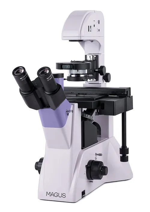
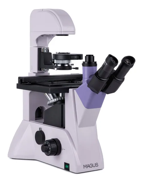

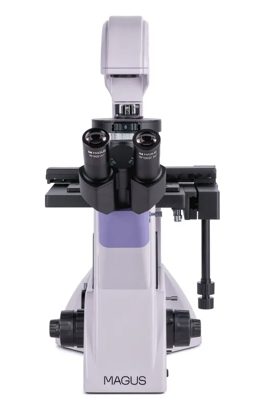


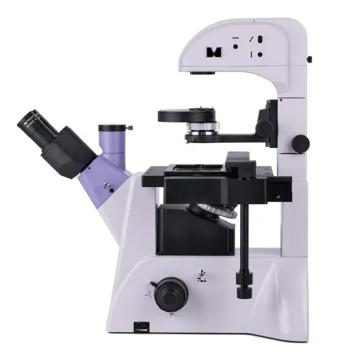


Magnification: 100–400x. Trinocular head, plan achromatic lenses, 30W halogen illuminator, phase contrast condenser, Köhler illumination
| Product ID | 82907 |
| Brand | MAGUS |
| Warranty | 5 years |
| EAN | 5905555018119 |
| Package size (LxWxH) | 49x34x73 cm |
| Shipping Weight | 19.5 kg |
MAGUS Bio V350 is a biological inverted microscope, which is used for studying stained and unstained samples in laboratory ware – Petri dishes, flasks, plates. The height of the dishes can be 55mm, and if you tilt the illuminator stand – up to 165mm. The bottom thickness of compatible ware is up to 1.2mm. The microscope, which utilizes the brightfield field and phase contrast microscopy techniques in the transmitted light, has a trinocular tube/side port camera for mounting a digital camera and monitor. It is used for routine laboratory and research work as well as teaching.
Optics
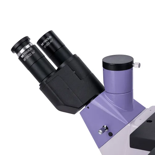
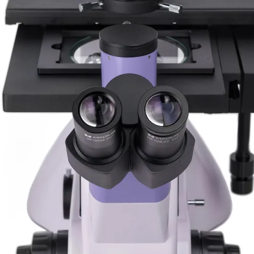
When assembling the microscope, you can select the position of the trinocular head: It can be rotated 180° to adjust the height of the exit pupil. Use the vertical tube to mount the monitor and the side port on the microscope body to mount a digital camera. The beam splitting on the body is 100/0 or 0/100 as well as 50/50 on the trinocular tube (when it is open) or 100/0 (when it is closed).
Four infinity plan achromatic objectives are included, one of which is a phase objective. Once four of them are installed in the revolving nosepiece, one slot is left free for an additional objective.
Illumination
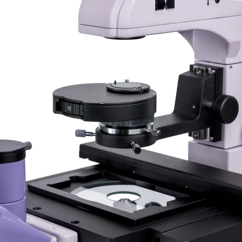
The phase-contrast condenser enables easy and convenient switching between microscopy techniques. It has four working positions: a free aperture for the brightfield technique as well as 10x, 20x, and 40x apertures for phase contrast using a suitable magnification objective.
The transmitted light source is a 30W halogen bulb that emits light of the color temperature that allows for comfortable work. It is brightness adjustable and is AC powered. The illumination system offers the use of the Köhler illumination to enhance image quality.
Stage and focusing mechanism
Four dish holders for the stage are included. They can be moved along the X and Y axes using a mechanical attachment, while the stage itself remains stationary. The holders are designed to hold labware of various sizes.
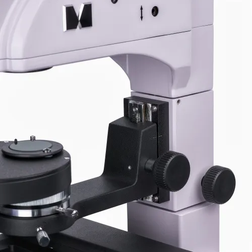
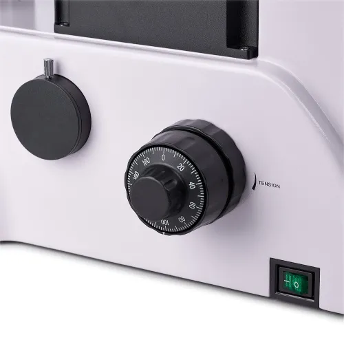
The focusing mechanism runs smoothly. The coarse focusing mechanism has a lock knob and tension adjustment. Fine focusing scale value: 2μm. The knobs are located at the base of the microscope so you can keep your hands in a more relaxed position while working.
Accessories
There are additional eyepieces, phase objectives, digital cameras, and calibration slides for the microscope to maximize the capabilities of the microscope.
Key features:
- For viewing samples in laboratory ware up to 55mm high and a bottom thickness up to 1.2mm
- If the illuminator stand is tilted, you can use dishes up to 165mm high
- Brightfield and phase-contrast techniques; phase-contrast condenser with a field diaphragm
- Stage with 4 dish holders of different sizes; mechanical attachment for moving the sample along two horizontal axes
- The microscope head rotates at 180°
- Two options for camera and monitor mounting: in the vertical tube and on the microscope body
- Transmitted light source – 30W halogen lamp powered by an AC power supply
- A wide range of optional accessories
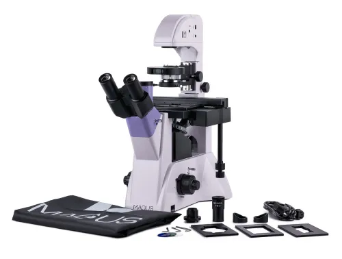
The kit includes:
- Stand with the built-in power supply, transmitted light source, focusing mechanism, stage, condenser mount, revolving nosepiece, and trinocular tube
- Phase-contrast condenser
- Trinocular head
- Infinity plan achromatic objective: PHP2 10x/0.25 phase WD 4.27mm
- Infinity plan achromatic objective: PL 10x/0.25 WD 4.27mm
- Infinity plan achromatic objective: PL 20x/0.40 WD 8.0mm
- Infinity plan achromatic objective: PL 40x/0.60 WD 3.5mm
- Eyepiece 10x/22mm with long eye relief (2 pcs.)
- Eyecup (2 pcs.)
- Centering telescope
- Round stage plate
- Mechanical attachment for moving the specimen
- Dish holder (4 pcs.)
- C-mount camera adapter
- Color filter
- AC power cord
- Dust cover
- User manual and warranty card
Available on request:
- 10x/22mm eyepiece with a scale
- 12.5x/14mm eyepiece (2 pcs.)
- 15x/15mm eyepiece (2 pcs.)
- 20x/12mm eyepiece (2 pcs.)
- 25x/9mm eyepiece (2 pcs.)
- Infinity plan achromatic objective: PL 4x/0.10 WD 21mm
- Infinity plan achromatic objective: PHP2 20x/0.40 phase WD 8.0mm
- Infinity plan achromatic objective: PHP2 40x/0.60 phase WD 3.5mm
- Digital camera
- Calibration slide
- LCD Monitor
| Product ID | 82907 |
| Brand | MAGUS |
| Warranty | 5 years |
| EAN | 5905555018119 |
| Package size (LxWxH) | 49x34x73 cm |
| Shipping Weight | 19.5 kg |
| Type | biological, light/optical |
| Microscope head type | trinocular |
| Head | Siedentopf, rotatable 180° |
| Head inclination angle | 45 ° |
| Magnification, x | 100 — 400 |
| Magnification, x (optional) | 40–500/600/800/1000 |
| Eyepiece tube diameter, mm | 30 |
| Eyepieces | 10х/22mm, eye relief: 10mm (*optional: 10x/22mm with scale, 12.5x/14; 15x/15; 20x/12; 25x/9) |
| Objectives | infinity plan achromatic: PL 10x/0.25, PL 20x/0.40, PL 40x/0.60, PHP2 10x/0.25 phase; parfocal distance 45mm (*optional: PL 4x/0.10, PHP2 20x/0.40 phase, PHP2 40x/0.60 phase) |
| Revolving nosepiece | for 5 objectives |
| Working distance, mm | 4.27 (10x); 8.0 (20х); 3.5 (40x) |
| Interpupillary distance, mm | 48 — 75 |
| Stage, mm | 227x208 |
| Stage moving range, mm | 77/114 |
| Stage features | fixed, with glass plate Ø118mm and mechanical attachment; dish holders: 86x129.5, Ø90mm; 34x77.5mm, Ø68.5mm; 57x82mm, Ø60mm; 29x77.5mm, Ø35mm |
| Condenser | NA 0.6, working distance: 55mm; phase contrast turret |
| Diaphragm | adjustable aperture diaphragm, adjustable iris field diaphragm |
| Focus | coaxial, coarse (with coarse focusing tension adjustment and a lock knob), and fine (0.002mm) |
| Illumination | halogen |
| Brightness adjustment | ✓ |
| Power supply | 220±22V, 50Hz, AC network |
| Light source type | 12V/30W |
| Light filters | yes |
| Operating temperature range, °C | 5...+35 |
| User level | experienced users, professionals |
| Assembly and installation difficulty level | complicated |
| Phase contrast device | phase contrast condenser (turret) with a free slot and phase annuli plates for 10x, 20x and 40x objectives; centering auxiliary microscope |
| Application | laboratory/medical |
| Illumination location | upper (transmitted light) |
| Research method | bright field, phase-contrast microscopy |
| Pouch/case/bag in set | dust cover |
We have gathered answers to the most frequently asked questions to help you sort things out
Find out why studying eyes under a microscope is entertaining; how insects’ and arachnids’ eyes differ and what the best way is to observe such an interesting specimen
Read this review to learn how to observe human hair, what different hair looks like under a microscope and what magnification is required for observations
Learn what a numerical aperture is and how to choose a suitable objective lens for your microscope here
Learn what a spider looks like under microscope, when the best time is to take photos of it, how to study it properly at magnification and more interesting facts about observing insects and arachnids
This review for beginner explorers of the micro world introduces you to the optical, illuminating and mechanical parts of a microscope and their functions
Short article about Paramecium caudatum - a microorganism that is interesting to observe through any microscope
I'm interested in the MAGUS Bio V350 Biological Inverted Microscope and have a question about its optical configuration. Specifically, could you confirm whether the microscope comes with an integrated tube lens for the side camera port? If so, could you please provide the focal length of that tube lens?
Thank you in advance for your prompt reply!
Best regards,
Pouya
Thank you for your query!
The imaging channel is lens-free. A single adapter from the microscope kit is mounted there if the camera matrix is large; or a 0.65x adapter for small matrixes should be purchased (C-mount MAGUS CFA065 Adapter No.: 83437). The camera can be attached to the adapter by screwing it on.