
 BG
BG  BY
BY  CY
CY  CZ
CZ  DE
DE  EE
EE  ES
ES  GR
GR  HU
HU  IS
IS  IT
IT  LT
LT  LV
LV  MY
MY  PL
PL  PT
PT  RO
RO  SK
SK  TR
TR  UA
UA  USA
USA MAGUS Bio V300 Biological Inverted Microscope
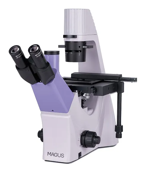
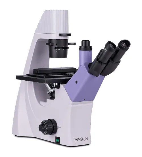
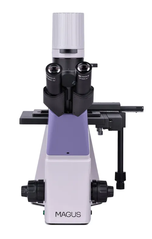
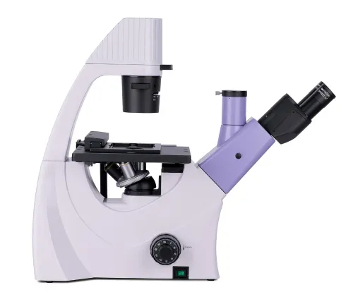
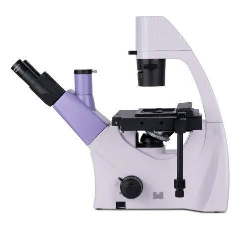
Magnification: 100–400x. Trinocular head, infinity plan achromatic objectives, 9W LED illuminator, condenser with a phase slider slot
| Product ID | 82906 |
| Brand | MAGUS |
| Warranty | 5 years |
| EAN | 5905555018089 |
| Package size (LxWxH) | 64x42x67 cm |
| Shipping Weight | 18 kg |
MAGUS Bio V300 is a biological microscope of an inverted design for studying samples in laboratory ware up to 70 mm high and with a bottom thickness up to 1.2 mm. The objects of study can be colonies of cells, tissue cultures, biological fluids, etc. The samples do not require mandatory pre-staining. The observations are made in transmitted light. Microscopy techniques: brightfield and phase contrast. The microscope is suitable for routine and research work, and teaching.
Optics
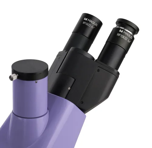
The microscope is equipped with a classic trinocular head, the trinocular tube is used for mounting a digital camera. The camera is not included in the set. The beam splitting is 50/50.
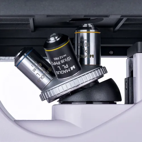
Four included or additional objectives can be mounted in the revolving nosepiece. All objectives are designed to be used with laboratory ware and have a long working distance. There are 3 plan achromatic objectives for brightfield and 1 plan achromatic objective for the phase contrast technique.
Illumination
The transmitted light source is a 9W LED. The high power of the LED makes the illumination of the working area bright enough to work with all objectives and with any microscopy technique. The brightness adjustment does not change the color temperature. The LED lifetime is about 50,000 hours without LED replacement.
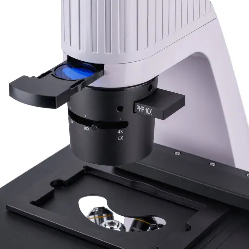
The condenser has a slot for a phase contrast slider (included). The phase contrast rings can be centered. The installation of the slider saves time when switching between the microscopy techniques.
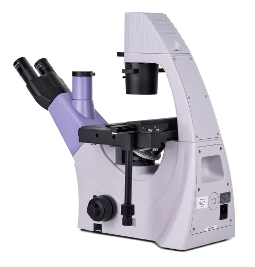
Stage and focusing mechanism
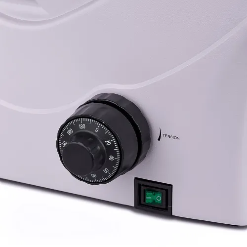
Fixed mechanical stage. It is equipped with a dish holder (three dish holders of different sizes are included). The dishes can be moved along the X and Y axes by a mechanical attachment. The coaxial coarse and fine focusing mechanism provides accurate and fast focusing. The coarse focusing has a lock mechanism and tension adjustment.
Accessories
The optional accessories line includes phase objectives, eyepieces, digital cameras, and calibration slides.
Key features:
- Brightfield and phase contrast microscopy techniques in the transmitted light; studying samples in laboratory ware
- Digital camera is mounted in the trinocular tube
- 9W LED transmitted light illuminator with adjustable brightness and a long lifetime
- Fixed stage with two-axis movement, 3 dish holders for different sizes included
- Optional accessories to improve microscope performance
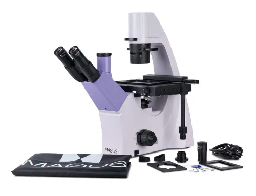
The kit includes:
- Stand with the built-in power supply, transmitted light source, focusing mechanism, stage, condenser with a slot for phase-contrast slider, and revolving nosepiece
- Trinocular head
- Infinity plan achromatic objective: PL 10x/0.25 WD 4.3mm
- Infinity plan achromatic objective: PL L20х/0.40 WD 8.0mm
- Infinity plan achromatic objective: PL L40х/0.60 WD: 3.5mm
- Infinity plan achromatic objective: PHP2 10x/0.25 phase WD 4.3mm
- Eyepiece 10x/22mm with long eye relief (2 pcs.)
- Eyecup (2 pcs.)
- Centering telescope
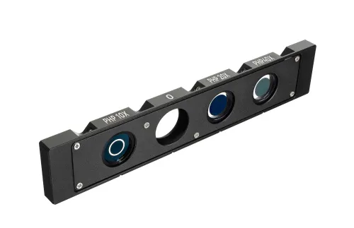
- Phase contrast slider with centering phase rings
- Mechanical attachment for moving the specimen
- Dish holder (3 pcs.)
- C-mount camera adapter
- Color filter
- AC power cord
- Dust cover
- User manual and warranty card
Available on request:
- 10x/22mm eyepiece with a scale
- 12.5x/14mm eyepiece (2 pcs.)
- 15x/15mm eyepiece (2 pcs.)
- 20x/12mm eyepiece (2 pcs.)
- 25x/9mm eyepiece (2 pcs.)
- Infinity plan achromatic objective: PL 4x/0.10 WD 21mm
- Infinity plan achromatic objective: PHP2 20x/0.40 phase WD 8.0mm
- Infinity plan achromatic objective: PHP2 40x/0.60 phase WD 3.5mm
- Digital camera
- Calibration slide
- LCD Monitor
| Product ID | 82906 |
| Brand | MAGUS |
| Warranty | 5 years |
| EAN | 5905555018089 |
| Package size (LxWxH) | 64x42x67 cm |
| Shipping Weight | 18 kg |
| Type | biological, light/optical |
| Microscope head type | trinocular |
| Head | Siedentopf |
| Head inclination angle | 45 ° |
| Magnification, x | 100 — 400 |
| Magnification, x (optional) | 40–500/600/800/1000 |
| Eyepiece tube diameter, mm | 30 |
| Eyepieces | 10х/22mm, eye relief: 10mm (*optional: 10x/22mm with scale, 12.5x/14; 15x/15; 20x/12; 25x/9) |
| Objectives | infinity plan achromatic: PL 10x/0.25, PL 20x/0.40, PL 40x/0.60, PHP2 10x/0.25 phase; parfocal distance 45mm (*optional: PL 4x/0.10, PHP2 20x/0.40 phase, PHP2 40x/0.60 phase) |
| Revolving nosepiece | for 4 objectives |
| Working distance, mm | 21.0 (4x); 4.3 (10x); 8.0 (20х); 3.5 (40x) |
| Interpupillary distance, mm | 48 — 75 |
| Stage moving range, mm | 77/112 |
| Stage features | fixed, with mechanical attachment for moving the specimen; dish holders: 29x77mm, Ø90mm; 34x77.5mm, Ø68.5mm; 57x82mm, Ø60mm |
| Condenser | NA 0.6, working distance: 70mm; with adjustable aperture diaphragm and a phase contrast slider slot |
| Diaphragm | adjustable aperture |
| Focus | coaxial, coarse (with coarse focusing tension adjustment and a lock knob), and fine (0.002mm) |
| Illumination | LED |
| Brightness adjustment | ✓ |
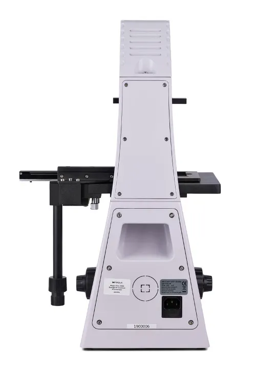
| Power supply | 220±22V, 50Hz, AC network |
| Light source type | 9W |
| Light filters | yes |
| Operating temperature range, °C | 5...+35 |
| Special features | phase contrast slider for a 10x objective with a centerable phase ring |
| User level | experienced users, professionals |
| Assembly and installation difficulty level | complicated |
| Application | laboratory/medical |
| Illumination location | upper (transmitted light) |
| Research method | bright field, phase-contrast microscopy |
| Pouch/case/bag in set | dust cover |
We have gathered answers to the most frequently asked questions to help you sort things out
Find out why studying eyes under a microscope is entertaining; how insects’ and arachnids’ eyes differ and what the best way is to observe such an interesting specimen
Read this review to learn how to observe human hair, what different hair looks like under a microscope and what magnification is required for observations
Learn what a numerical aperture is and how to choose a suitable objective lens for your microscope here
Learn what a spider looks like under microscope, when the best time is to take photos of it, how to study it properly at magnification and more interesting facts about observing insects and arachnids
This review for beginner explorers of the micro world introduces you to the optical, illuminating and mechanical parts of a microscope and their functions
Short article about Paramecium caudatum - a microorganism that is interesting to observe through any microscope
