
 BG
BG  BY
BY  CY
CY  CZ
CZ  DE
DE  EE
EE  ES
ES  GR
GR  HU
HU  IS
IS  IT
IT  LT
LT  LV
LV  MY
MY  PL
PL  PT
PT  RO
RO  SK
SK  TR
TR  UA
UA  USA
USA MAGUS Bio D260T LCD Biological Digital Microscope
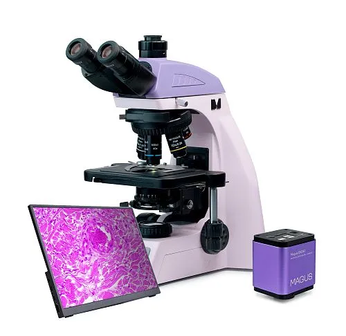
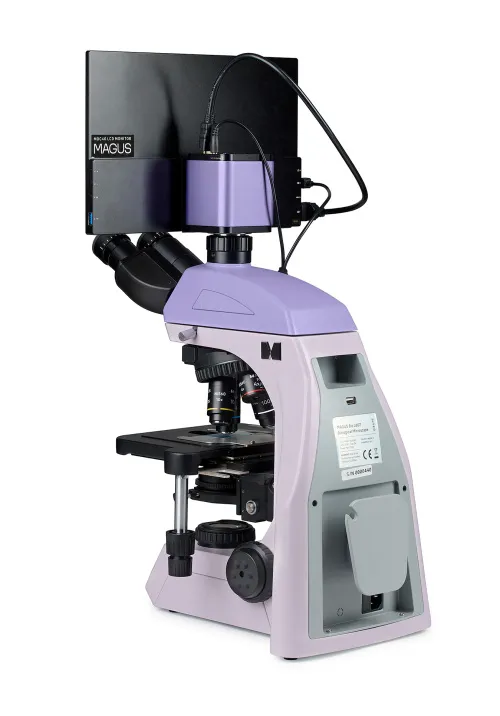
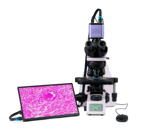


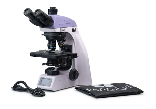


With a camera and a monitor. Magnification: 40–1000x. Trinocular microscope head, coded revolving nosepiece, plan achromatic objectives, 3W LED illuminator, and intelligent lighting control system
| Product ID | 83284 |
| Brand | MAGUS |
| Warranty | 5 years |
| EAN | 5905555019918 |
| Package size (LxWxH) | 47x32x67 cm |
| Shipping Weight | 15.05 kg |
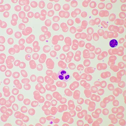
The microscope is suitable for observing transparent and translucent biological samples, such as smears and cross sections using the brightfield microscopy technique in transmitted light. Optional accessories allow for using darkfield, phase-contrast, fluorescence, and polarization microscopy. The coded revolving nosepiece maintains a comfortable brightness level when the objectives are changed. The microscope's intelligent lighting control system improves comfort and speed of the researcher’s everyday work. Smart features help students ease into the profession and gain the professional experience they need. The microscope is suitable for laboratory work, research, and education purposes. The microscope is equipped with an 8MP camera and three video interfaces: HDMI, USB 2.0, and WLAN. It can be connected simultaneously to several devices: monitor, laptop, and desktop computer. The software complements the system with analysis and documentation functions. The monitor in a digital microscope has 4K resolution.
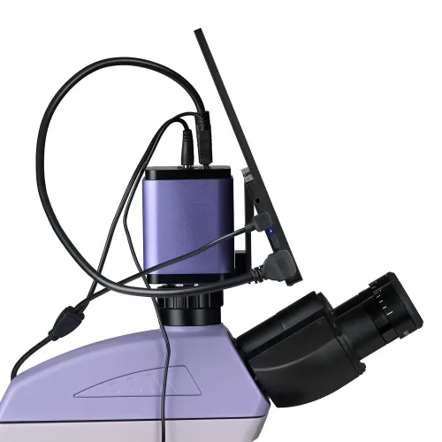
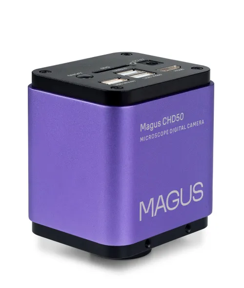
Digital camera
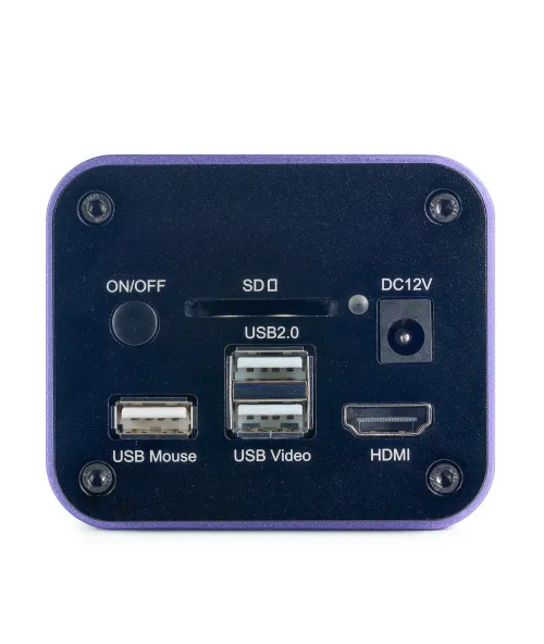
MAGUS CHD50 is a digital autofocus HDMI camera supporting three video interfaces. The camera features an 8MP sensor and produces realistic images in 4K resolution (3840x2160px) automatically switching between 4K and Full HD depending on the resolution of the monitor. The camera uses an HDMI interface to connect directly to a TV, monitor, or projector. In this mode, the camera operates autonomously without a PC. The HDMI interface provides a high and stable transfer rate from the camera to the external screen. You can connect it to a PC via WLAN or USB2.0. Video is recorded at 30fps. The camera combines high FPS and high bandwidth HDMI and, therefore, videos are vivid with no freezes or gaps between frames. At maximum resolution, the image is well-detailed, moving objects are visible without any bugs, and object movement is displayed without delays.
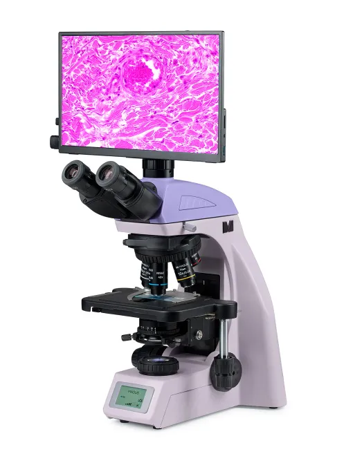
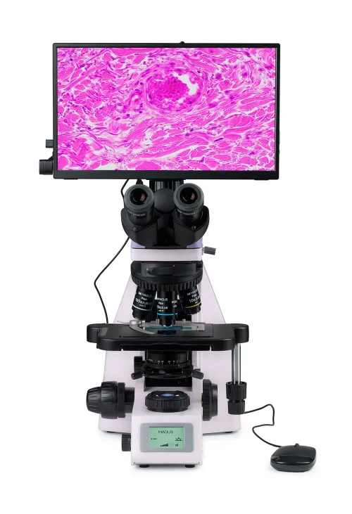
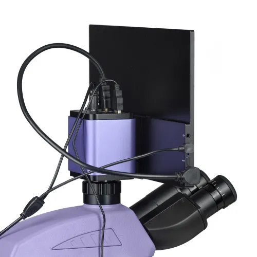
Monitor
The MAGUS MCD40 Monitor is designed to use a visualization system of the MAGUS microscope. It connects to a microscope-mounted camera to display a real-time image. It is compatible with MAGUS HDMI cameras operating in 4К resolution. The screen diagonal is 13.3 inches. The IPS sensor delivers a bright image with large viewing angles: If you look at the monitor at an angle, there is no color distortion. The display can be placed on a folding stand on a table or mounted directly on the camera, whichever is more convenient for the user.
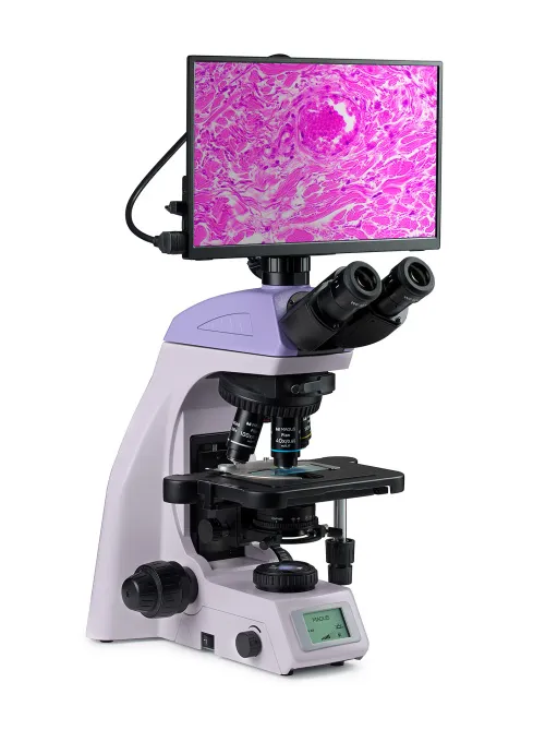
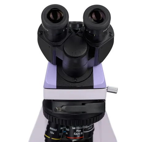
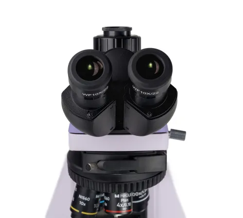
Microscope head
Trinocular, with infinity-corrected optics. The eyepiece tubes are 360° rotatable. The user can adjust the eyepiece height to suit their individual stature. The digital camera is mounted in the trinocular tube. The beam splitting ratio is fixed at 50/50. The vertical tube provides 0.55x magnification. When paired with the included 1x adapter, the final magnification at the camera is 0.55x. When selecting a camera and calculating the resolution and sensor size, use the final magnification factor of 0.55x. The basic configuration includes 10x/22mm eyepieces with diopter adjustment and eye relief to work with glasses. Flat rubber eyecups protect eyeglasses from scratches.
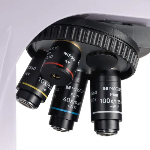
Revolving nosepiece and objectives
The coded revolver for five objectives is oriented toward the interior: The user can see the objective inserted into the optical path, and the space above the stage is free. An additional objective can also be installed in the free slot in order to achieve extra magnification. The parfocal distance is 60mm. The 40x objective with its extended working distance of 1.5mm provides additional protection for the front lens against immersion oil contact when rotating the nosepiece. Above the revolver, there is a slot with a plug for the analyzer.
Maintaining comfortable brightness levels when switching magnifications
The objectives of different magnifications transmit light with different levels of intensity, and so each time you change objectives, the brightness of the light must be adjusted. Switching from a higher to a lower magnification objective causes eye fatigue, as the image brightness in the eyepieces increases sharply. The MAGUS Bio D260T LCD, which is equipped with intelligent brightness control, solves this problem. The microscope remembers the brightness for each objective that the user has selected and automatically sets this brightness when turning the nosepiece. Intelligent control reduces the time required to adjust brightness. MAGUS Bio D260T LCD increases user comfort and saves time even when the given work requires frequent magnification changes.
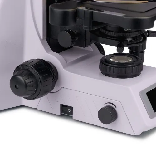
Focusing mechanism
Coaxial coarse and fine focusing knobs. The coarse focusing knob is located on the left side. Fine focusing knobs are located on both sides, the right knob having special finger indentations. The ring on the left side adjusts the tension of the coarse focusing travel. The user adjusts the comfortable tension for work.
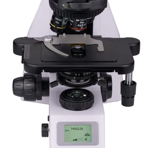
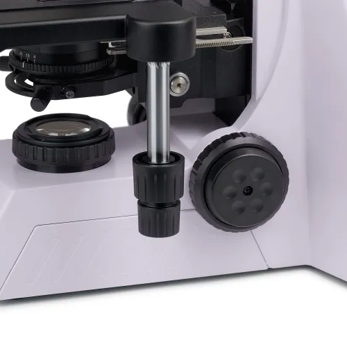
Stage
The stage has no X-axis rack and pinion, which improves ergonomics. The belt-driven mechanism allows for smooth movement of the specimen. The specimen holder is secured with two screws and can be easily removed during manual scanning. The long stage control handle ensures user comfort: The hand rests on the table with no strain.
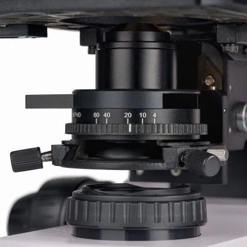
Condenser
The condenser is height-adjustable and can be centered. Mounting type: dovetail. The condenser ring adjusts the iris aperture diaphragm. The condenser body has a marking for the magnification of the objectives, and the ring has an indicator marker. To achieve contrast on each objective, it is recommended to rotate the ring so that the index marker matches the magnification marking of the objective being used. The condenser has a slot with a plug for a darkfield slider or a phase contrast slider. The installation of a slider saves time when changing the observation method.
Light source
The transmitted light illuminator contains a 3W LED. The color temperature does not change when you adjust the brightness. The LED lifetime is 50,000 hours.
Köhler illumination in transmitted light
The Köhler illumination improves the image quality of the observed sample: Each objective achieves maximum resolution and the field of view is illuminated evenly without darkening at the edges. The object of study is in sharp focus and the image artifacts are removed.
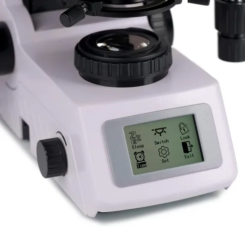
Status LCD screen
The LCD screen on the base of the microscope displays the objective magnification, brightness and color temperature of the light source, and operating mode (“sleep” and “eco”). Using the screen and two knobs, the microscope user adjusts the brightness, locks the brightness adjustment, and sets the sleep mode and auto-off timer.
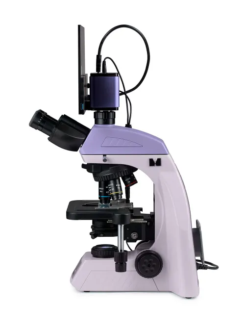
Ergonomic design
Physical discomfort causes fatigue and reduces productivity. The ergonomic design of the microscope plays an important role in everyday scientific research. MAGUS Bio D260T LCD provides user comfort during work. By rotating the microscope head, the user selects a point of view height so as not to strain their back and neck. Focusing knobs are located at the bottom of the body. The user does not need to strain their hands. Thanks to the smooth movement of the mechanism, the user can effortlessly focus on the object. The microscope is operated with minimal hand movement, since the long stage control handle and fine focusing knob are located in the same working area. The microscope is equipped with a special carrying handle. The design for the hidden placement of the power cord improves the workplace esthetic and safety of carrying the microscope as well as simplifies storage of the device.
Accessories
There is a line of accessories designed specifically for this microscope. Optional objectives provide additional magnification. Eyepieces extend the magnification range of the microscope. Optional eyepieces help you maximize the potential of the objective that you use most often. The phase contrast device, epi-fluorescence attachment, darkfield slider, and polarizer/analyzer set offer more contrast techniques so that you can study specimens that are invisible in brightfield. A calibration slide is used to measure objects and can be combined with the eyepiece with a scale or with the camera software.
Alternative models
MAGUS Bio D260T LCD is an alternative for the following models: Carl Zeiss Primostar 3, Leica DM 1000, Leica DM 1000LED, Leica DM 2000, Leica DM 2000LED, Olympus CX 33.
Microscope features:
- Observations of transparent and translucent samples in brightfield in transmitted light
- The trinocular head with a vertical tube for installing a digital camera and height adjustment to adjust to the observer; light beam splitting 50/50
- Coded revolving nosepiece: The brightness of the light source is set automatically depending on the selected objective
- Transmitted light illuminator is energy-saving 3W LED with a lifetime of up to 50,000 hours
- Transmitted Köhler illumination, centered and height-adjustable condenser with objective magnification markings
- Smart lighting control system: automatic brightness selection, dimming lock, timer auto-off, LCD operating status screen
- Stage without an X-axis positioning rack and with a long control handle for convenient operation
- Ergonomic stand with a carrying handle and the hidden placement of the power cord
- Wide range of compatible optional accessories
Camera features:
- The camera operates autonomously via an HDMI interface without connecting to a computer or installing software. It can also connect to a computer via WLAN and USB 2.0 interfaces
- Autofocus in HDMI mode makes it easy to adjust image sharpness
- Auto switching between 4K and Full HD depending on the monitor resolution
- 30fps with any video output interface for observing moving objects, recording video, and moving the sample without jerkiness or delays
- SONY Exmor/Starvis color CMOS backlit sensor provides low noise level and high light sensitivity even in low-light conditions. You will get clearer, brighter, and more color-saturated images
- The software with the functions of photography, video recording, editing, and external monitor display as well as linear and angular measurement
Monitor features:
- 4K resolution provides highly detailed, clear, and realistic images
- The IPS sensor produces a bright and saturated picture with wide vertical and horizontal viewing angles
- Can be mounted on the camera or table to suit the user’s preferences
Package:
- MAGUS CHD50 digital camera (digital camera, HDMI cable (1.5m), USB 3.0 cable (2m), USB mouse, 32GB SD memory card, 12V/1A (Euro) power adapter, USB flash drive with drivers and software, user manual, and warranty card)
- MAGUS MCD40 LCD Monitor
- Stand with a built-in power supply, transmitted light source, focusing mechanism, stage, condenser mount, and revolving nosepiece
- Abbe condenser
- Trinocular microscope head
- Plan achromatic objective Plan 4x/0.10 ∞/–, parfocal distance: 60mm
- Plan achromatic objective Plan 10x/0.25 ∞/–, parfocal distance: 60mm
- Plan achromatic objective Plan 40x/0.65 ∞/0.17, parfocal distance: 60mm
- Plan achromatic objective Plan 100x/1.25 oil ∞/0.17 (spring-loaded), parfocal distance: 60mm
- 10х/22mm eyepiece with long eye relief and diopter adjustment (2 pcs.)
- Eyecup (2 pcs.)
- C-mount adapter
- Microscope power cord
- Dust cover
- User manual and warranty card
Available on request:
- 10х/22mm eyepiece with a scale
- 10x/22mm eyepiece with a reticle
- 10x/22mm eyepiece with crosshairs
- 12.5х/16mm eyepiece (2 pcs.)
- 15х/16mm eyepiece (2 pcs.)
- 20х/12mm eyepiece (2 pcs.)
- 30х/8mm eyepiece (2 pcs.)
- Plan achromatic objective Plan 2х/0.06 ∞/–, parfocal distance: 60mm
- Plan achromatic objective Plan 20х/0.40 ∞/0.17, parfocal distance: 60mm
- Plan achromatic objective Plan 60х/0.80 ∞/0.17, parfocal distance: 60mm
- Plan apochromatic objective Plan Apo 60х/1.42 oil ∞/0.17, parfocal distance: 60mm
- Plan achromatic objective Plan 100х/1.10 ∞/0.17 water, parfocal distance: 60mm
- Set of plan semi-apochromatic fluorescence objectives: 4x, 10x, 20x, 40x, 100x (oil); ∞/0.17; parfocal distance 60mm
- Phase contrast device: set of phase sliders, set of phase objectives, auxiliary centering telescope
- Phase contrast device: phase contrast condenser, set of phase objectives, auxiliary centering telescope
- Darkfield slider
- Dry darkfield condenser NA 0.7–0.9
- Oil darkfield condenser NA 1.3–1.26
- Epi-fluorescence attachment with LED light source, fluorescence filter cubes, and power cable
- Polarizer/analyzer set
- Digital camera
- Calibration slide
- Set of color filters (blue, green, yellow, frosted glass)
- C-mount adapter
| Product ID | 83284 |
| Brand | MAGUS |
| Warranty | 5 years |
| EAN | 5905555019918 |
| Package size (LxWxH) | 47x32x67 cm |
| Shipping Weight | 15.05 kg |
| Type | biological, light/optical |
| Microscope head type | trinocular |
| Head | Gemel head (Siedentopf, 360° rotation), with 50/50 distribution of the luminous flux |
| Head inclination angle | 30 ° |
| Magnification, x | 40 — 1000 |
| Magnification, x (optional) | 40–1250/1500/2000 |
| Eyepiece tube diameter, mm | 30 |
| Eyepieces | 10x/22, long eye relief (*optional: 10x/22 with scale, 10x/22 with reticle, 10x/22 with crosshair, 12.5x/17.5, 15x/16, 20x/12) |
| Objectives | plan achromatic, infinity corrected: 4x/0.10; 10x/0.25; 40x/0.65; 100xs/1.25 oil (*optional: 20x/0.40); parfocal height 60mm |
| Revolving nosepiece | 5 objectives, coded |
| Working distance, mm | 30 (4x); 10.2 (10x); 1.5 (40x); 0.2 (100xs) |
| Interpupillary distance, mm | 47 — 78 |
| Stage, mm | 230x150 |
| Stage moving range, mm | 78/54 |
| Stage features | two-axis mechanical stage, without a positioning rack |
| Eyepiece diopter adjustment, diopters | ±5D on each eyepiece |
| Eyepiece diopter adjustment | ✓ |
| Condenser | center- and height-adjustable Abbe condenser NA 1.25 with adjustable aperture diaphragm and slot with plug for darkfield and phase contrast sliders; dovetail mount |
| Diaphragm | adjustable aperture diaphragm, adjustable iris field diaphragm |
| Focus | coaxial, coarse (30mm, 37.7mm/circle, with tension adjustment mechanism), and fine (0.002mm, 0.2mm/circle) |
| Illumination | LED |
| Brightness adjustment | ✓ |
| Power supply | 100–240V, 50/60Hz, AC network |
| Light source type | 3W LED |
| Operating temperature range, °C | 0...+70 |
| Additional | automatic brightness adjustment when switching objectives, eco mode, sleep mode, status display on LCD screen |
| Ability to connect additional equipment | darkfield condenser (dry or oil), darkfield slider, phase contrast device (condenser and objectives), phase contrast device (slider and objectives), polarization devices (polarizer and analyzer), reflected light illuminator |
| User level | experienced users, professionals |
| Application | laboratory/medical |
| Illumination location | lower |
| Research method | bright field |
| Pouch/case/bag in set | dust cover |
| Sensor | Sony Exmor/Starvis CMOS |
| Color/monochrome | color |
| Megapixels | 8 |
| Maximum resolution, pix | 3840x2160 (4K UHD) |
| Sensor size | 1/1.8" (7.68x4.32mm) |
| Pixel size, μm | 2x2 |
| Auto focus | ✓ |
| Interface connectors | HDMI 1.4, WLAN, USB2.0 |
| Memory card | SD up to 32GB |
| Light sensitivity | 505mV with 1/30s |
| Signal/noise ratio | 0.15mV at 1/30s |
| Exposure time | 0.04ms–1000ms |
| Video recording | ✓ |
| Frame rate, fps at resolution | 30@3840x2160 (HDMI), 30@3840x2160 (USB), 30@3840x2160 (WLAN) |
| Place of installation | trinocular tube, eyepiece tube instead of an eyepiece |
| Image format | *.jpg, *.tif |
| Spectral range, nm | 380–650 (built-in IR filter) |
| Shutter type | ERS (electronic rolling shutter) |
| Software | HDMI: built-in; USB: MAGUS View |
| System requirements | Windows 8/10/11 (32bit and 64bit), Mac OS X, Linux, up to 2.8GHz Intel Core 2 or higher, minimum 4GB RAM, USB2.0 port, RJ45, 19" or larger display |
| Mount type | C-mount |
| Body | CNC aluminum alloy |
| Camera power supply | DC adapter 12V, 1A |
| Camera operating temperature range, °С | -10...+50 |
| Operating humidity range, % | 30 — 80 |
| Type of matrix | IPS |
| Display diagonal, inch | 13.3 |
| Display resolution, px | 3840x2160 (4K) |
| Aspect ratio | 16:9 |
| Brightness, cd/m² | 400 |
| Number of displayed colors | 16.7 m |
| Contrast | 1000:1 |
| Horizontal/vertical viewing angle, ° | 178/178 |
| Size of the visible screen area (WxH), mm | 295x165 |
| Pixel pitch (WxH), mm | 0.154x0.154 |
| Frequency of optical source, Hz | 60 |
| Type of matrix backlight | LED |
| LED backlight lifetime, h | 50000 |
| Interface | HDMI |
| Operating temperature range, °C | -15...+55 |
| Operating humidity range, % | 10 — 90 |
| Power supply | AC 110–220V, DC 5–12V/1A (Type-C) |
| Power consumption, W | 12 (max.) |
We have gathered answers to the most frequently asked questions to help you sort things out
Find out why studying eyes under a microscope is entertaining; how insects’ and arachnids’ eyes differ and what the best way is to observe such an interesting specimen
Read this review to learn how to observe human hair, what different hair looks like under a microscope and what magnification is required for observations
Learn what a numerical aperture is and how to choose a suitable objective lens for your microscope here
Learn what a spider looks like under microscope, when the best time is to take photos of it, how to study it properly at magnification and more interesting facts about observing insects and arachnids
This review for beginner explorers of the micro world introduces you to the optical, illuminating and mechanical parts of a microscope and their functions
Short article about Paramecium caudatum - a microorganism that is interesting to observe through any microscope
