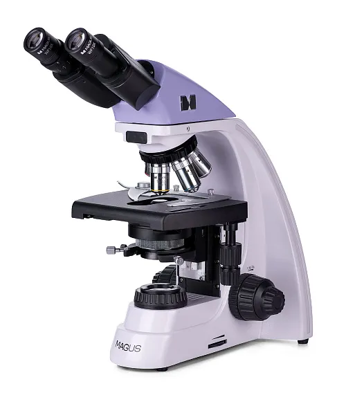
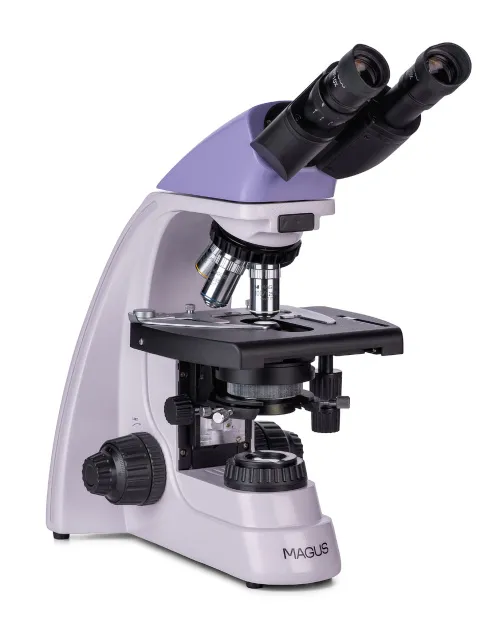
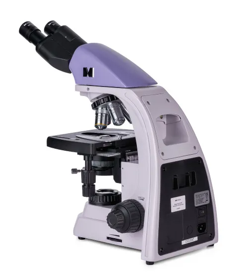
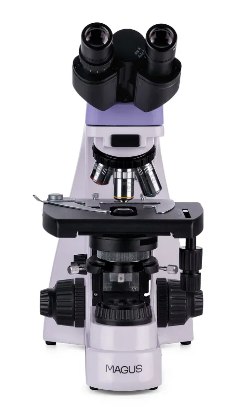
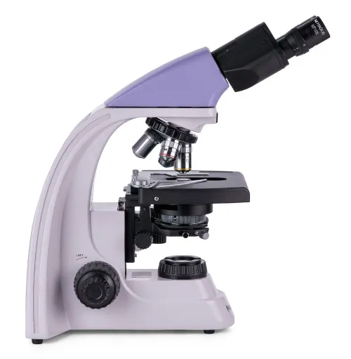
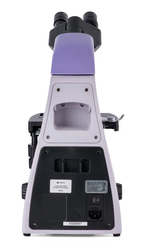
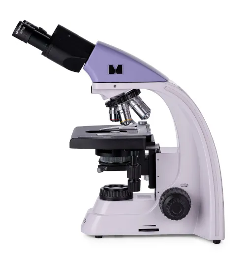
MAGUS Bio 230B Biological Microscope
Magnification: 40–1000x. Binocular head, achromatic objectives, 30W halogen bulb
| Product ID | 82892 |
| Brand | MAGUS |
| Warranty | 5 years |
| EAN | 5905555017921 |
| Package size (LxWxH) | 43x27x63 cm |
| Shipping Weight | 10 kg |
The MAGUS Bio 230B microscope is designed for laboratory and scientific research in order to observe biological specimens such as thin sections or smears. The transmitted light microscopy technique (brightfield) is used for sample analysis. If necessary, the microscope can be equipped with additional accessories for darkfield, polarized light, and phase contrast techniques. The microscope can be applied in such areas of research as pharmaceutical industry, medicine, biotechnology, environmental protection, forensics, and agriculture.
Optics
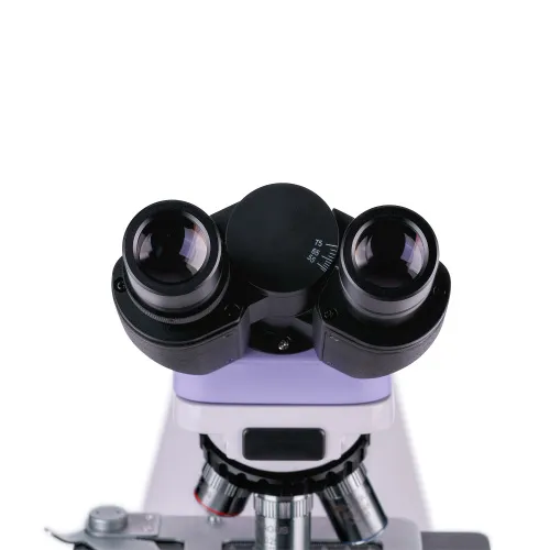
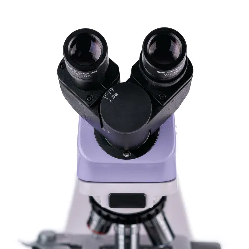
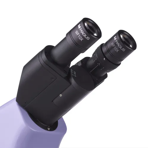
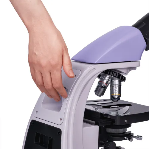
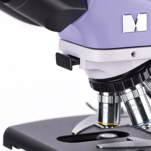
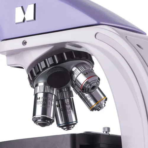
The microscope has a binocular head. The observation tubes can be rotated 360°, thereby allowing the user to adjust the position to fit their height. There is a diopter adjustment ring on the left tube. The revolving nosepiece has 5 slots, and it is oriented toward the interior – the user can see the objectives inserted into the optical path and, therefore, the space above the stage is free. Four achromatic objectives are included in the kit, and the fifth slot can be used to mount an optional objective.
The optics are infinity corrected. The basic microscope magnification ranges from 40 to 1000x. With additional eyepieces, it can be extended up to 1500, 1600, and 2000x. If necessary, a digital camera (not included) can be mounted into the eyepiece tube.
Illumination
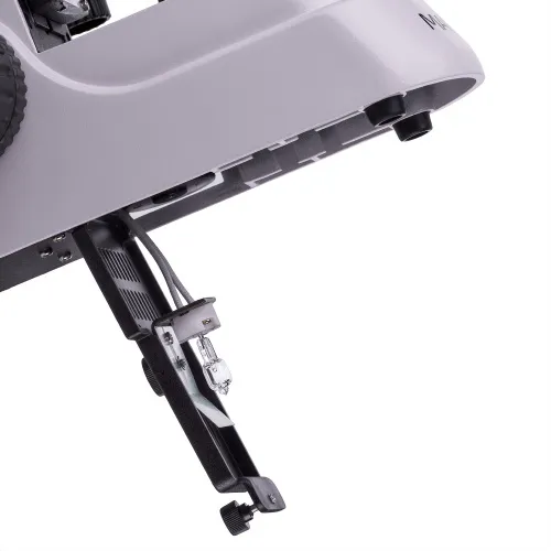
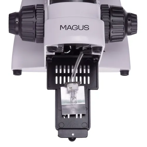
The light source is a halogen bulb located at the microscope base. The warm lighting reduces eye strain and is ideal for long hours of working with the microscope. The 30W halogen lamp enables bright and high-contrast images to be produced with any type of objective and any microscopy technique (including darkfield and phase contrast).
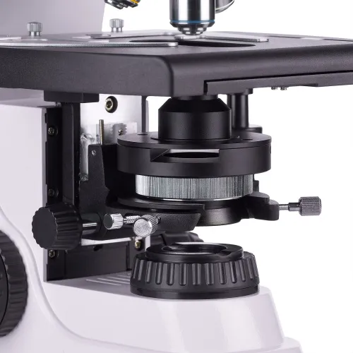
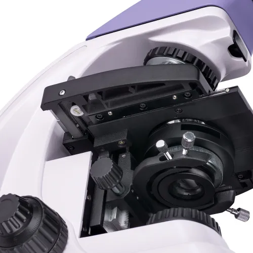
The Abbe condenser can be centered, and its position is height-adjustable. A phase contrast or darkfield slider can be installed in a special slot, thereby allowing you to easily switch between microscopy techniques. The field diaphragm makes it possible to set up the Köhler illumination.
Stage and focusing mechanism
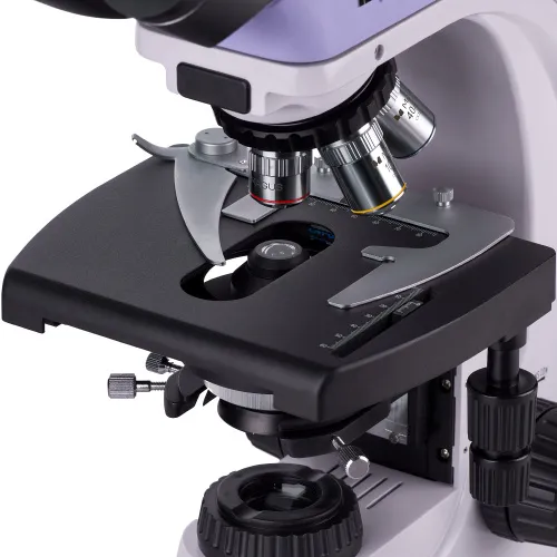
The stage does not have a positioning rack, which makes it easier to work with the microscope. A mechanical attachment, which can be removed, if necessary, ensures the smooth movement of the specimen across the stage.
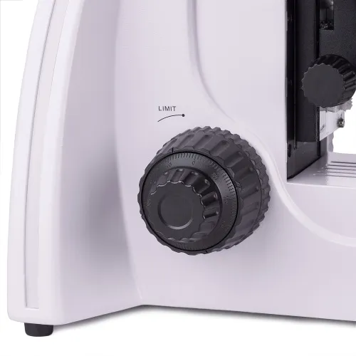
The coarse and fine focusing knobs are coaxial (on the same axis) and located on both sides at the lower part of the microscope. Such positioning of the focusing knobs enables you to rest your hands freely on the table and sit comfortably for long periods of work. The focusing adjustment is smooth and requires no further effort. The coarse focusing has a lock knob and focus tension adjustment.
Accessories
Additional equipment, such as objectives, eyepieces, phase contrast devices, darkfield condensers, polarizing devices, digital cameras, or other accessories, will expand the capabilities of the microscope. They are designed for MAGUS Bio 230B and available on request.
Key features:
- Infinity achromatic optics
- Binocular head, the observation tubes can be rotated 360°, diopter adjustment on the left tube
- Revolving nosepiece for 5 objectives, a stage without a positioning rack
- Coarse and fine focusing knobs, coarse focusing tension adjusting knob, coarse focusing lock knob
- Condenser with a slot for a phase or darkfield slider, field diaphragm for Köhler illumination
- 30W halogen bulb (transmitted light source), brightness-adjustable, powered by the AC power supply
- Option to install additional accessories to expand the capabilities of the microscope
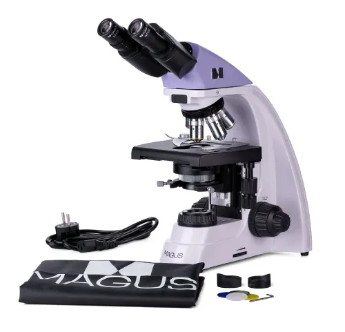
The kit includes:
- Base with a power input, transmitted light source, focusing mechanism, stage, condenser mount, and revolving nosepiece
- Abbe condenser
- Binocular head
- Infinity achromatic objective: 4x/0.10
- Infinity achromatic objective: 10x/0.25
- Infinity achromatic objective: 40x/0.65 (spring-loaded)
- Infinity achromatic objective: 100x/1.25 oil (spring-loaded)
- Eyepiece 10x/18mm with a long eye relief (2 pcs.)
- Eyecup (2 pcs.)
- Filter (4 pcs.)
- Bottle of the immersion oil
- AC power cord
- Dust cover
- User manual and warranty card
Available on request:
- 10x/20mm eyepiece with a scale
- 15x/11mm eyepiece (2 pcs.)
- 16x/11mm eyepiece (2 pcs.)
- 20x/11mm eyepiece (2 pcs.)
- Infinity achromatic objective: 20x/0.40
- Infinity plan achromatic objective: 20х/0.40
- Infinity plan achromatic objective: 60x/0.80 (spring-loaded)
- Phase-contrast device
- Phase slider
- Darkfield condenser NA 0.9
- Oil darkfield condenser NA 1.36–1.25
- Darkfield slider
- Polarization devices
- Digital camera
- Calibration slide
- C-mount adapter
- LCD Monitor
| Product ID | 82892 |
| Brand | MAGUS |
| Warranty | 5 years |
| EAN | 5905555017921 |
| Package size (LxWxH) | 43x27x63 cm |
| Shipping Weight | 10 kg |
| Type | biological, light/optical |
| Microscope head type | binocular |
| Head | Gemel head (Siedentopf, 360° rotation) |
| Head inclination angle | 30 ° |
| Magnification, x | 40 — 1000 |
| Magnification, x (optional) | 40–1500/1600/2000 |
| Eyepiece tube diameter, mm | 23.2 |
| Eyepieces | 10х/18mm, eye relief: 10mm (*optional: 10х/20mm with a scale, 15x/11mm, 16x/11mm; 20x/11mm) |
| Objectives | infinity achromatic: 4x/0.10; 10x/0.25; 40xs/0.65; 100xs/1.25 (oil); parfocal distance 45mm (*optional: 20x/0.40; 60xs/0.80) |
| Revolving nosepiece | for 5 objectives |
| Working distance, mm | 18.89 (4x); 5.95 (10x); 0.775 (40xs); 0.36 (100xs); 2.61/8.80 (20x); 0.46 (60хs) |
| Interpupillary distance, mm | 48 — 75 |
| Stage, mm | 180x150 |
| Stage moving range, mm | 75/50 |
| Stage features | two-axis mechanical stage, without a positioning rack |
| Eyepiece diopter adjustment, diopters | ±5 (on the left tube) |
| Condenser | Abbe condenser, N.A. 1.25, center-adjustable, height-adjustable, adjustable aperture diaphragm, a slot for a darkfield slider and phase contrast slider, dovetail mount |
| Diaphragm | adjustable aperture diaphragm, adjustable iris field diaphragm |
| Focus | coaxial, coarse focusing (21mm, 39.8mm/circle, with a lock knob and tension adjusting knob) and fine focusing (0.002mm) |
| Illumination | halogen |
| Brightness adjustment | ✓ |
| Power supply | 220±22V, 50Hz, AC network |
| Light source type | 12V/30W halogen bulb, G4 |
| Light filters | yes |
| Ability to connect additional equipment | phase contrast device (condenser and objectives), darkfield condenser (dry or oil), polarization devices (polarizer and analyzer) |
| User level | experienced users, professionals |
| Assembly and installation difficulty level | complicated |
| Application | laboratory/medical |
| Illumination location | lower |
| Research method | bright field |
| Pouch/case/bag in set | dust cover |
| Weight, kg | 8 |
| Dimensions, mm | 200x436x400 |
| Camera operating temperature range, °С | 5...+35 |
We have gathered answers to the most frequently asked questions to help you sort things out
Find out why studying eyes under a microscope is entertaining; how insects’ and arachnids’ eyes differ and what the best way is to observe such an interesting specimen
Read this review to learn how to observe human hair, what different hair looks like under a microscope and what magnification is required for observations
Learn what a numerical aperture is and how to choose a suitable objective lens for your microscope here
Learn what a spider looks like under microscope, when the best time is to take photos of it, how to study it properly at magnification and more interesting facts about observing insects and arachnids
This review for beginner explorers of the micro world introduces you to the optical, illuminating and mechanical parts of a microscope and their functions
Short article about Paramecium caudatum - a microorganism that is interesting to observe through any microscope
You recommanded me this device to mesure perforation holes of leather product from 0.9 MM up to 2.1 MM with 0.01 Precision.
My question is how to take picture of the hole and how to mesure with that precision? is it an additionnal software ar it's already intergrated inside this microscope?
Second question how to fix the leather part under the objective, this material have an average thickness reaching 2 MM.
thank you in advance
Noomen
Thanks for your query.
I double checked with our technical support, they replied that for your purposes it is better to use 82910_MAGUS Stereo 9T with both top and bottom lights on at the same time (top light is brighter and bottom light is darker), then you can get a good image of the leather hole.
Here is the product link: https://eu.levenhuk.com/catalogue/microscopes/magus-stereo-9t-stereoscopic-microscope/?oid=52994
Both recommended microscopes do not have built-in software.
And the 2 mm thick sample is perfectly fixed in the microscope clamps.
Hope this helps!

