BG
BG  BY
BY  CY
CY  CZ
CZ  DE
DE  EE
EE  ES
ES  GR
GR  HU
HU  IS
IS  IT
IT  LT
LT  LV
LV  MY
MY  PL
PL  PT
PT  RO
RO  SK
SK  TR
TR  UA
UA  USA
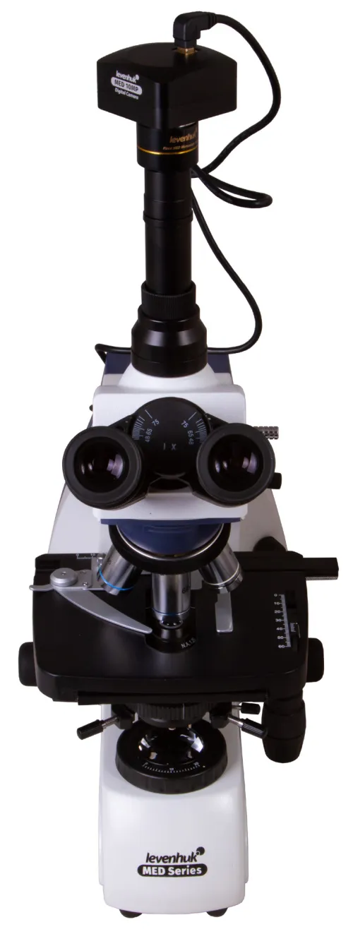
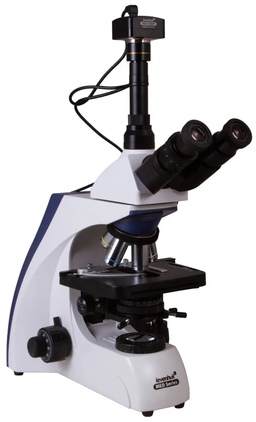
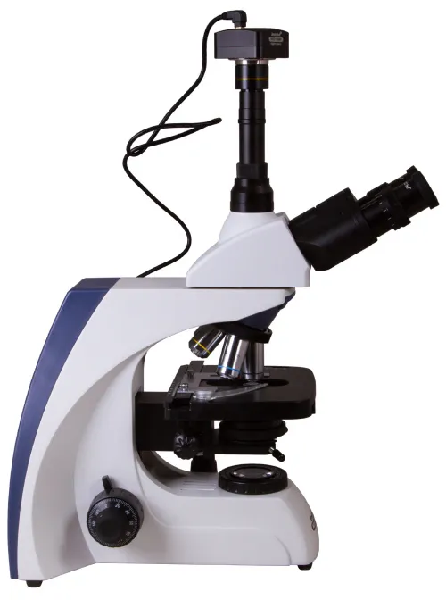
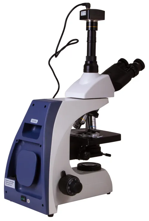
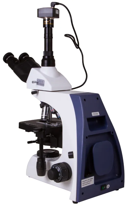
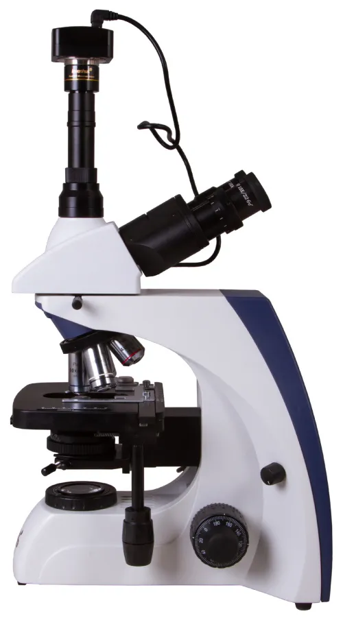
USA Levenhuk MED D35T Digital Trinocular Microscope
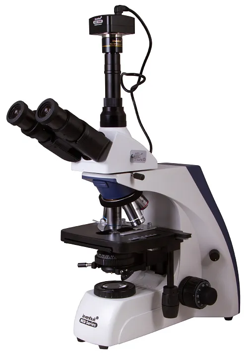
Magnification: 40–1000x. Trinocular head, 10MP digital camera, infinity-corrected plan achromatic objective lenses, an Abbe condenser with an iris diaphragm
| Product ID | 74002 |
| Brand | Levenhuk, Inc., USA |
| Warranty | lifetime |
| EAN | 5905555005041 |
| Package size (LxWxH) | 62x36x25 cm |
| Shipping Weight | 10.92 kg |
The Levenhuk MED D35T Digital Microscope for scientific research combines the capabilities of a classic biological model and a microscope for taking digital photos and videos. It comes with a 10MP camera that is connected to a computer and allows for observing an image on its screen in real-time mode. This microscope is excellent for a university department, research center, or clinical and diagnostics laboratory.
Plan achromatic infinity-corrected optics
The microscopes in the Levenhuk MED 35 series are equipped with an infinity-corrected optical system that is used in professional and high-class microscopes. This system includes Infinity PlanAchromat objectives and allows for obtaining clear, high contrast images with high level of flatness.
One of the most important features of an infinity-corrected optical system is that it allows for installing any additional parts in the optical path between objective lenses and an ocular tube. The additional parts include polarizers and epi-fluorescence light. All in all, modular design and simple operation make Levenhuk MED 35 optimal microscopes for using in different kinds of microscopy and working in hematological, histological, microbiological, and other laboratories.
The Levenhuk MED 35 series includes scientific microscopes for working in a laboratory
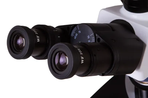
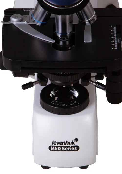
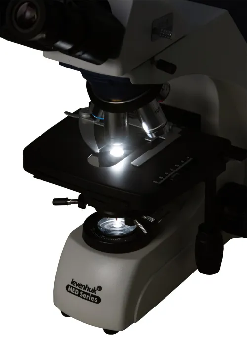
A trinocular head has a binocular visual unit and a vertical tube inclined at 30° for installing a digital camera in it. There is a beam splitter. The head is 360° rotatable, which is convenient if the sample should be observed by several researchers.
Capabilities of the optical system
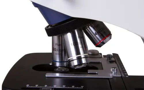
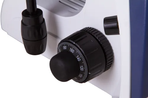
Scientific and research work with Levenhuk MED D35T microscope is practical and precise. The image is formed by wide-field 10x eyepieces with diopter adjustment and five plan achromatic objectives with various magnification. Plan achromatic optics flatten the field of view, reduce chromatic aberrations, and improve color rendering. You can conduct highly detailed observations at magnifications of 40x to 1000x. The objective lenses with 40x, 60x, and 100x magnifications are equipped with spring-loaded protective frames; the 100x objective lens is used for oil immersion observations. The sharpness is adjusted with the coarse and fine focusing adjustment knobs.
Convenient work with microscope slides
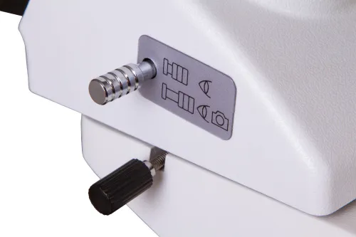
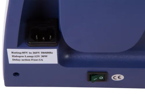
The stage is moveable along two axes and equipped with a mechanical scale. Underneath, there is a halogen light (30W) with adjustable brightness. An Abbe condenser with an iris diaphragm is used for directing an optical beam. Three optical filters are included in the kit. You can also set up Köhler illumination. The light is powered by an AC power supply.
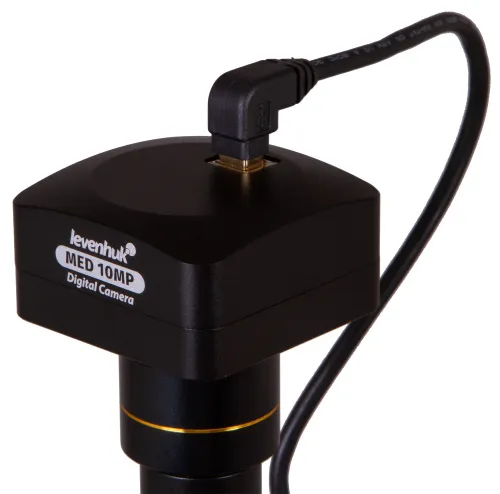
A 10M digital camera for capturing photos and videos
A 10MP sensor in a digital camera (included in the kit) allows for obtaining high-resolution pictures. Moreover, recording smooth videos with high frame rate is available. A camera transmits an image to a computer screen online. That makes work in a laboratory easier and more practical as well as reduces eyestrain and tension in the rotator cuff, as there is no necessity to look through the eyepieces. Special software (on an included CD) allows for the simple processing of recorded images. The software allows for adjusting size, image brightness and contrast, exposure time and white balance, calibrating a camera and objective lenses, measuring specimens or structures (several measurement units are available). A digital camera is powered and connected to a computer via a USB cable.
Scientific work with Levenhuk MED D35T Microscope provides for precise observations, allows for using modern technologies, and conducting practical extended observations.
Features:
- Magnification: 40x to 1000x
- Trinocular head with a beam splitter, wide-field eyepieces
- Plan achromatic optics with an anti-fungal coating
- Halogen light with adjustable brightness
- Setting up Köhler illumination is available
- Powered by an AC power supply
- 10MP digital camera is included
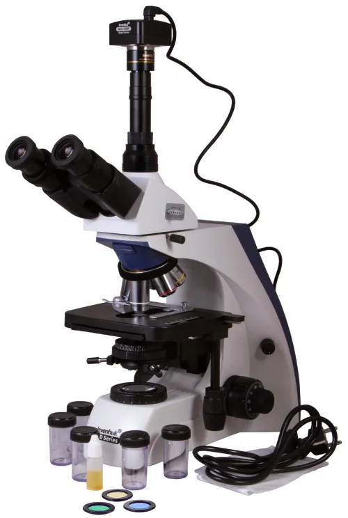
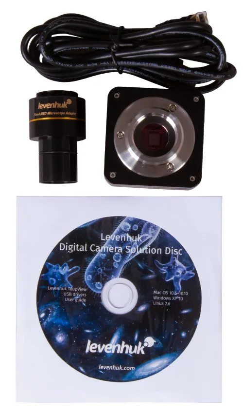
The kit includes:
- Microscope base with a stand
- 360° rotatable trinocular head
- Infinity-corrected plan achromatic objective lenses: 4x, 10x, 40xs, 60xs, 100xs (oil) with an anti-fungal coating
- Wide-field eyepieces: WF10x/22mm with an anti-fungal coating (2 pcs)
- Abbe condenser N.A. 1.25 with an iris diaphragm
- Filters: blue, green, yellow
- Bottle of immersion oil
- Fuse (2 pcs)
- Power cord for microscope
- Dust cover
- 10MP digital camera
- Camera adapter
- Camera mount
- USB cable for connecting and powering a digital camera
- Software CD and drivers
- User manual and lifetime warranty
Caution:
Please refer to the specifications table for the correct mains voltage and never attempt to plug a 110V device into 220V outlet and vice versa without using a converter. Remember that mains voltage in the U.S. and Canada is 110V and 220–240V in most European countries.
Some things you can see under a microscope:


Levenhuk MED D35T Digital Trinocular Microscope is also compatible with other Levenhuk digital cameras (additional cameras are purchased separately). Levenhuk cameras are installed in the eyepiece tube instead of an eyepiece. This microscope is also compatible with any other digital microscope cameras.
| Product ID | 74002 |
| Brand | Levenhuk, Inc., USA |
| Warranty | lifetime |
| EAN | 5905555005041 |
| Package size (LxWxH) | 62x36x25 cm |
| Shipping Weight | 10.92 kg |
| Type | biological, light/optical, digital |
| Microscope head type | trinocular |
| Optics material | optical glass with anti-fungal coating |
| Head | 360 ° rotatable, with switching (dividing) luminous flux |
| Head inclination angle | 30 ° |
| Magnification, x | 40 — 1000 |
| Eyepiece tube diameter, mm | 23.2 mm (third vertical tube), 30mm (binocular head) |
| Eyepieces | WF10x/22mm (2 pcs.) |
| Objectives | infinity-corrected plan achromatic objective lenses: 4x, 10x, 40xs, 60xs, 100xs (oil) |
| Revolving nosepiece | for 5 objectives |
| Interpupillary distance, mm | 48 — 75 |
| Stage, mm | 180x160 |
| Stage moving range, mm | 80/50 (movement in horizontal (X and Y) directions) |
| Coarse focusing travel, mm | 20 |
| Stage features | mechanical double-layer |
| Eyepiece diopter adjustment, diopters | ±5 |
| Condenser | Abbe N. A. 1.25 with an iris diaphragm |
| Diaphragm | iris |
| Focus | coaxial, coarse (0.5 mm) and fine (0.002 mm), with rack and pinion |
| Body | metal |
| Illumination | halogen |
| Brightness adjustment | ✓ |
| Power supply | 100–240V |
| Light source type | 12V/30W, 85–230V AC |
| Light filters | blue, green, yellow |
| Additional | Köhler illumination, collector lens |
| User level | experienced users, professionals |
| Assembly and installation difficulty level | complicated |
| Application | laboratory/medical |
| Illumination location | lower |
| Research method | bright field |
| Pouch/case/bag in set | dust cover |
| Camera specifications | |
| Megapixels | 10 |
| Sensor element | 1/2.3" |
| Pixel size, μm | 1.67x1.67 |
| Video recording | yes |
| Image format | *.jpg, *.bmp, *.png, *.tif and others |
| Video format | output: *.wmv, *.avi, *.h264 (Windows 8 and later), *h265 (Windows 10 and later) |
| Spectral range, nm | 380–650 |
| White balance | automatic, manual |
| Exposure control | 0.4–2000μs |
| Sensitivity, V/lux-sec@550nm | 0.31 |
| Frame rate | 3.3@3584x2748 11@1792x1374 38@896x684 |
| Dynamic range, dB | 65.2 |
| Usage location | the third 23.2mm ocular tube of the microscope |
| Software, drivers | Levenhuk |
| Programmable options | brightness, image size, shutter time |
| Output | USB 2.0 |
| System requirements | Windows 8/10/11 (32bit and 64bit), Mac OS X, Linux, up to 2.8GHz Intel Core 2 or higher, minimum 2GB RAM, USB 2.0 port, CD-ROM |
| Camera power supply | via USB cable |
and downloads
We have gathered answers to the most frequently asked questions to help you sort things out
Find out why studying eyes under a microscope is entertaining; how insects’ and arachnids’ eyes differ and what the best way is to observe such an interesting specimen
Read this review to learn how to observe human hair, what different hair looks like under a microscope and what magnification is required for observations
Learn what a numerical aperture is and how to choose a suitable objective lens for your microscope here
Learn what a spider looks like under microscope, when the best time is to take photos of it, how to study it properly at magnification and more interesting facts about observing insects and arachnids
This review for beginner explorers of the micro world introduces you to the optical, illuminating and mechanical parts of a microscope and their functions
Short article about Paramecium caudatum - a microorganism that is interesting to observe through any microscope