BG
BG  BY
BY  CY
CY  CZ
CZ  DE
DE  EE
EE  ES
ES  GR
GR  HU
HU  IS
IS  IT
IT  LT
LT  LV
LV  MY
MY  PL
PL  PT
PT  RO
RO  SK
SK  TR
TR  UA
UA  USA
USA Levenhuk MED 40T Trinocular Microscope
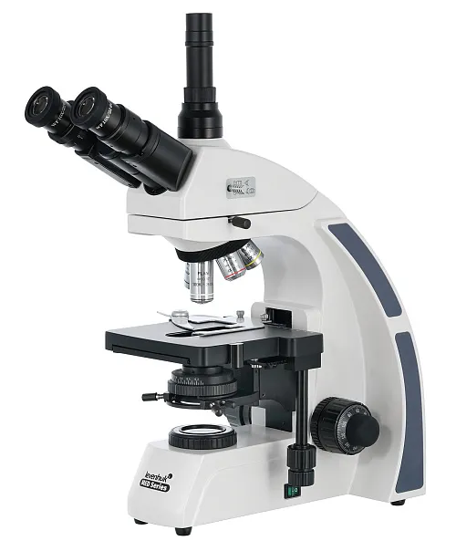
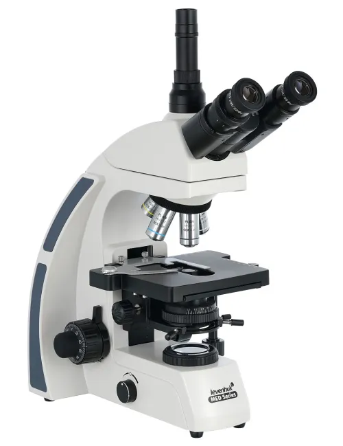
Magnification: 40x to 1000x. Trinocular head, infinity-corrected plan achromatic objective lenses, an Abbe condenser with an iris diaphragm
| Product ID | 74005 |
| Brand | Levenhuk, Inc., USA |
| Warranty | lifetime |
| EAN | 5905555005126 |
| Package size (LxWxH) | 50x44x28 cm |
| Shipping Weight | 9.98 kg |
The Levenhuk MED 40T is a trinocular microscope with an infinity-corrected plan achromatic optical system that can be used for professional 40x to 1000x microscopic studies. The microscope produces a clear and high-contrast image without any distortions and enables studying small structures on a fully flattened field. It will be a perfect choice for a professional of a clinical diagnostic laboratory, employee of a scientific research center, or educational institution.
Plan achromatic infinity-corrected optics
The microscopes in the Levenhuk MED 40 series are equipped with an infinity-corrected optical system that is used in professional and high-class microscopes. This system includes Infinity PlanAchromat objectives and allows for obtaining clear, high contrast images with a high level of flatness.
One of the most important features of an infinity-corrected optical system is that it allows for installing any additional parts in the optical path between objective lenses and an ocular tube. The additional parts include polarizers and epi-fluorescence light. All in all, modular design and simple operation make Levenhuk MED 40 optimal microscopes for use in different kinds of microscopy and working in hematological, histological, microbiological, and other laboratories.
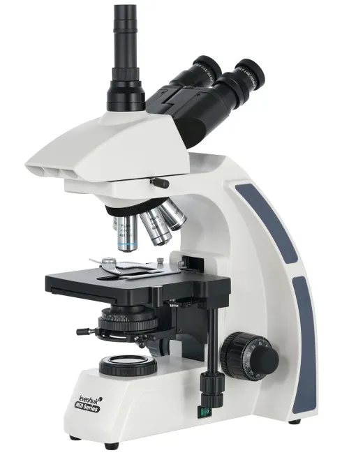
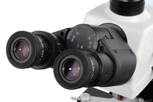
A rotatable head with a beam splitter
The trinocular head enables you to simultaneously observe samples in a traditional way and display pictures on an external screen online. The head consists of a binocular visual part and a separate tube where a camera can be fitted (the camera is not included in the kit). The visual module is inclined by 30°, which prevents tensing your neck muscles. The tube for a digital camera is vertical. For comfortable group observations, the head can be rotated by 360°. There is a beam splitter.
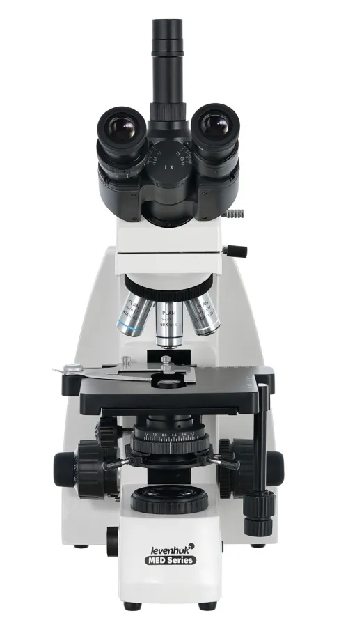
Capabilities of the optical system
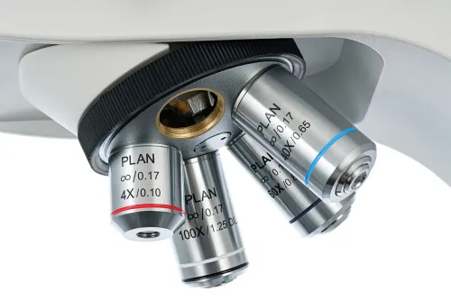
The optical system consists of two wide-field eyepieces and five plan achromatic objective lenses. Observations can be made by a standard or immersion method (a 100x objective lens is used for oil immersion). Eyepieces have 10x magnification and diopter adjustment. Three objectives with the maximum magnification are fitted with spring rims that protect the optics from incidental damage. All optic surfaces of accessories are protected with anti-fungal coating. Coarse and fine focusing is available.
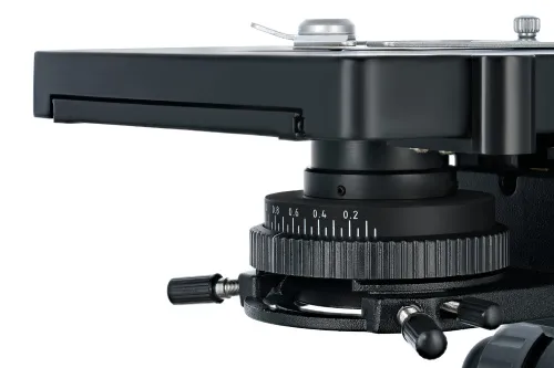
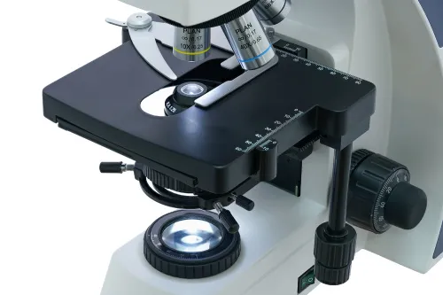
Convenient work with microscope slides
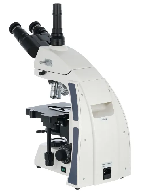
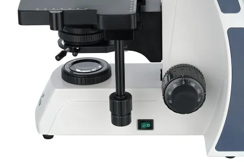
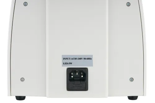
Under the optical system, there is a stage with a mechanical scale. Underneath, there is an Abbe condenser with an iris diaphragm. Specimens are illuminated from the bottom with a bright 5W LED. Köhler illumination is available.
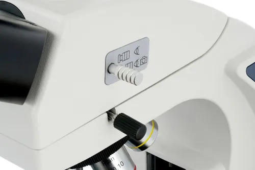
The light requires an AC power supply. The light brightness is adjustable.
Features:
- Infinity-corrected plan achromatic optics
- Trinocular head with a beam splitter, magnification range of 40x to 1000x
- Bright LED light
- Köhler illumination is available
- Powered by an AC power supply, adjustable light brightness
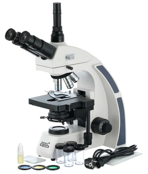
The kit includes:
- Microscope base with a stand
- 360° rotatable trinocular head
- Infinity-corrected plan achromatic objective lenses: 4x, 10x, 40xs, 60xs, 100xs (oil) with anti-fungal coating
- Wide-field eyepieces: WF10x/22mm with anti-fungal coating (2 pcs)
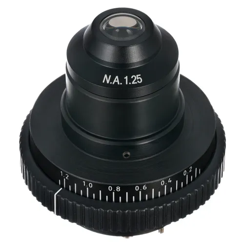
- Abbe condenser N.A. 1.25 with an iris diaphragm
- Filters: blue, green, yellow
- Bottle of immersion oil
- Fuse (2 pcs)
- Power cord for microscope
- Dust cover
- Camera mount
- User manual and lifetime warranty
Caution:
Please refer to the specifications table for the correct mains voltage and never attempt to plug a 110V device into 220V outlet and vice versa without using a converter. Remember that mains voltage in the U.S. and Canada is 110V and 220–240V in most European countries.
Some things you can see under a microscope:


Levenhuk MED 40T Trinocular Microscope is also compatible with other Levenhuk digital cameras (additional cameras are purchased separately). Levenhuk cameras are installed in the eyepiece tube instead of an eyepiece. This microscope is also compatible with any other digital microscope cameras.
| Product ID | 74005 |
| Brand | Levenhuk, Inc., USA |
| Warranty | lifetime |
| EAN | 5905555005126 |
| Package size (LxWxH) | 50x44x28 cm |
| Shipping Weight | 9.98 kg |
| Type | biological, light/optical |
| Microscope head type | trinocular |
| Optics material | optical glass with anti-fungal coating |
| Head | 360 ° rotatable, with switching (dividing) luminous flux |
| Head inclination angle | 30 ° |
| Magnification, x | 40 — 1000 |
| Eyepiece tube diameter, mm | 23.2 mm (third vertical tube), 30mm (binocular head) |
| Eyepieces | WF10x/22mm (2 pcs.) |
| Objectives | infinity-corrected plan achromatic objective lenses: 4x, 10x, 40xs, 60xs, 100xs (oil) |
| Revolving nosepiece | for 5 objectives |
| Interpupillary distance, mm | 48 — 75 |
| Stage, mm | 180x150 |
| Stage moving range, mm | 75/50 (movement in horizontal (X and Y) directions) |
| Coarse focusing travel, mm | 48 |
| Stage features | mechanical double-layer |
| Eyepiece diopter adjustment, diopters | ±5 |
| Condenser | Abbe N. A. 1.25 with an iris diaphragm |
| Diaphragm | iris |
| Focus | coaxial, coarse (0.5 mm) and fine (0.002 mm), with rack and pinion |
| Body | metal |
| Illumination | LED |
| Brightness adjustment | ✓ |
| Power supply | 100–240V |
| Light source type | 5W, 85–230 V AC |
| Light filters | blue, green, yellow |
| Additional | Köhler illumination, collector lens |
| User level | experienced users, professionals |
| Assembly and installation difficulty level | complicated |
| Application | laboratory/medical |
| Illumination location | lower |
| Research method | bright field |
| Pouch/case/bag in set | dust cover |
We have gathered answers to the most frequently asked questions to help you sort things out
Find out why studying eyes under a microscope is entertaining; how insects’ and arachnids’ eyes differ and what the best way is to observe such an interesting specimen
Read this review to learn how to observe human hair, what different hair looks like under a microscope and what magnification is required for observations
Learn what a numerical aperture is and how to choose a suitable objective lens for your microscope here
Learn what a spider looks like under microscope, when the best time is to take photos of it, how to study it properly at magnification and more interesting facts about observing insects and arachnids
This review for beginner explorers of the micro world introduces you to the optical, illuminating and mechanical parts of a microscope and their functions
Short article about Paramecium caudatum - a microorganism that is interesting to observe through any microscope