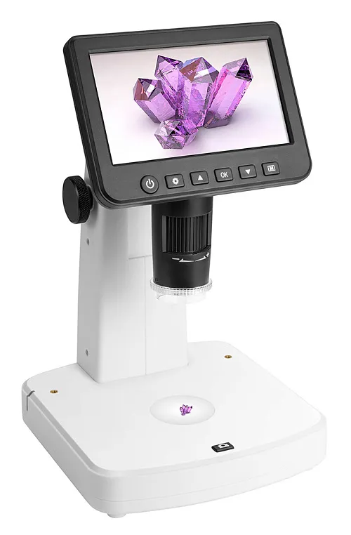
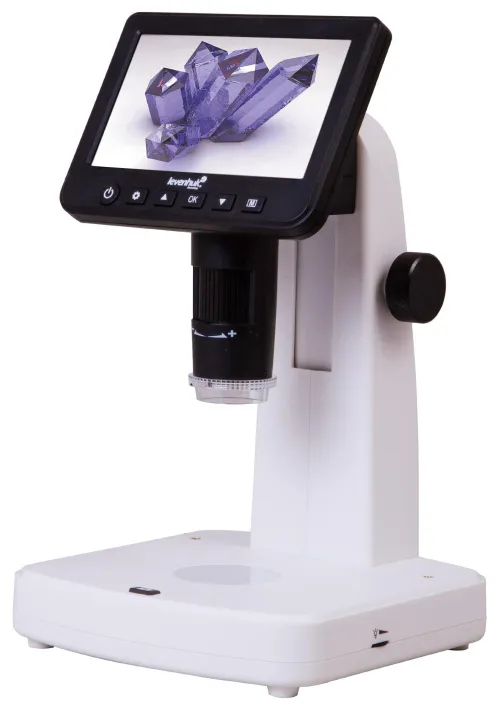
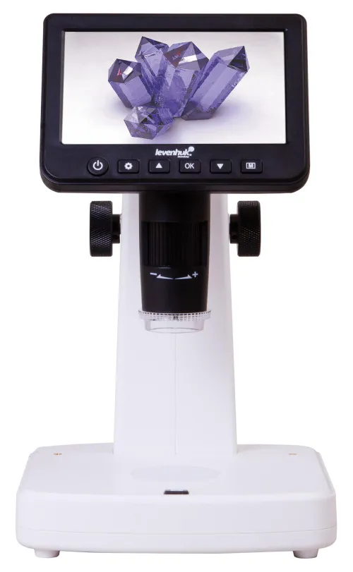
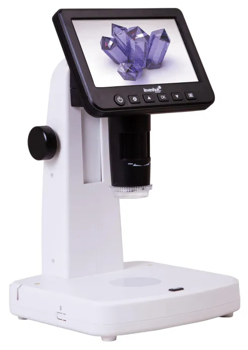
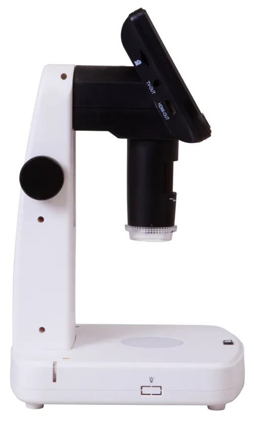
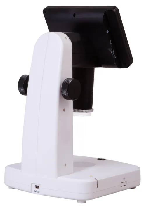
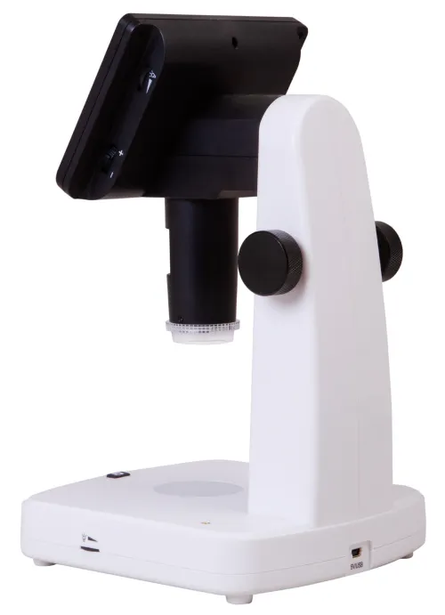
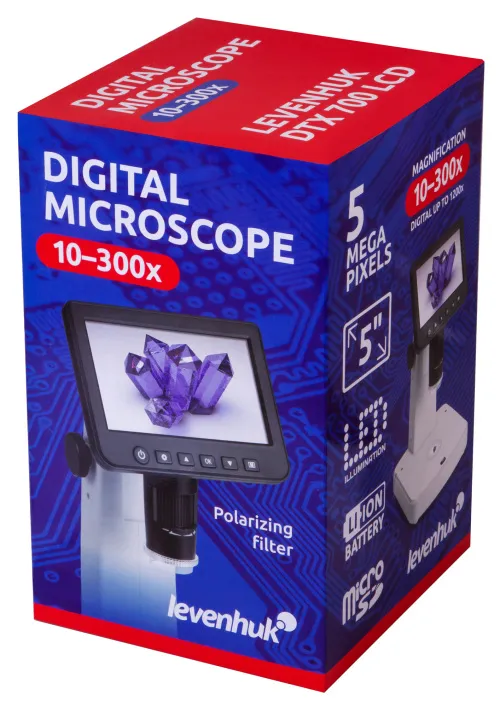
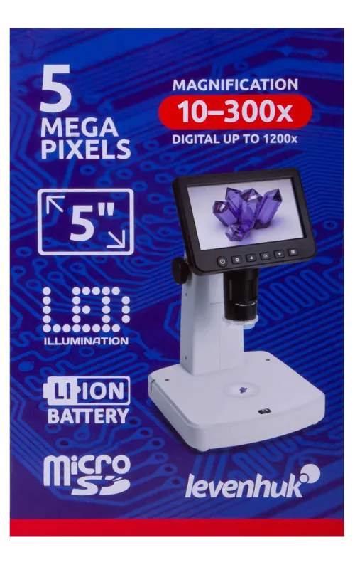
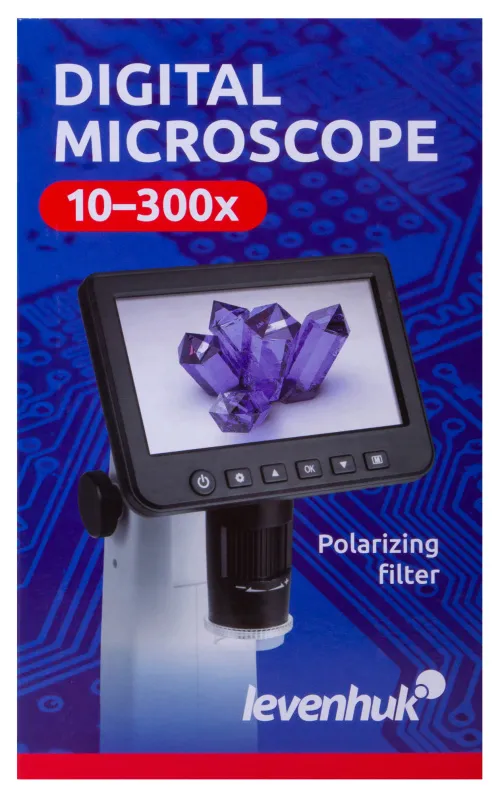
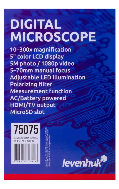
Levenhuk DTX 700 LCD Digital Microscope
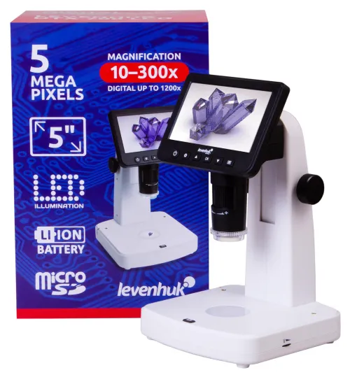
Magnification: 10–300x (with a digital zoom: 10–1200x). Digital USB microscope with an LCD screen and a 5M camera
| Product ID | 75075 |
| Brand | Levenhuk, Inc., USA |
| Warranty | lifetime |
| EAN | 5905555004389 |
| Package size (LxWxH) | 31x20x19 cm |
| Shipping Weight | 1.52 kg |
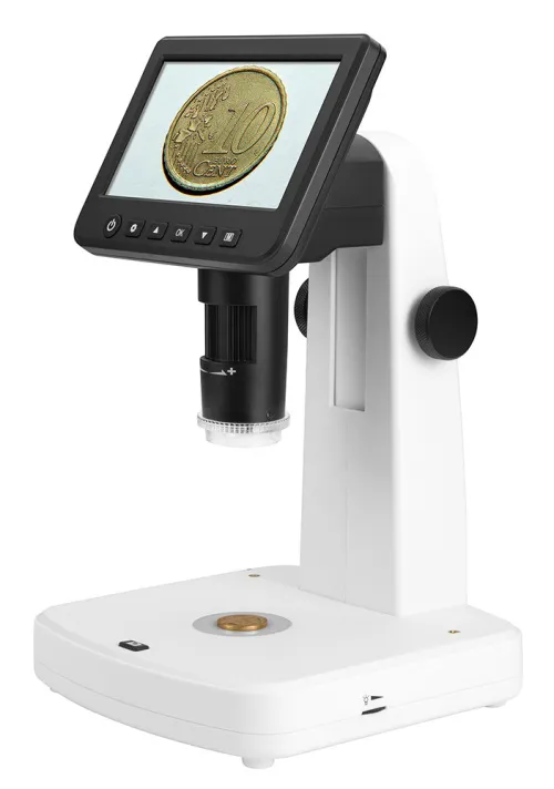
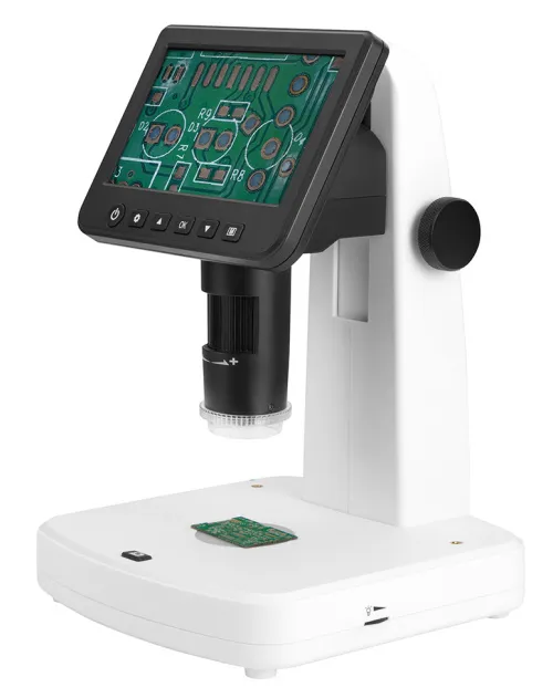
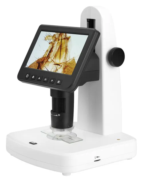
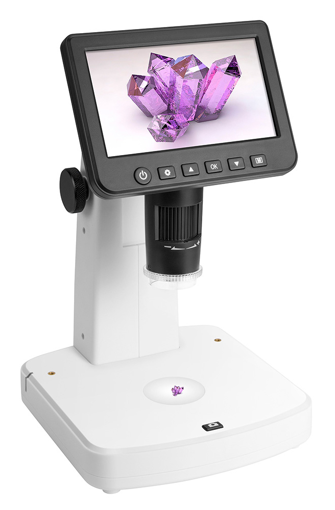
Levenhuk DTX 700 LCD Digital Microscope is suitable for working with jewelry pieces, electronic plates, minerals, coins, and sections of metals. This microscope is also useful for home use, e.g. for studying insects and plants. Levenhuk DTX 700 LCD is equipped with an LCD screen, that reduces eyestrain during lengthy work, and it is more comfortable than looking through an eyepiece of a standard microscope.
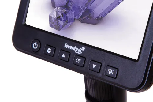
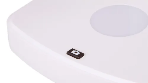
An image is formed by a 5M digital camera. The camera provides 10–300x magnification, and an additional zoom allows for observing objects at up to 1200x magnification. A colorful and detailed image is transmitted to a built-in LCD screen. In addition, a built-in camera allows for capturing images and shooting videos. Control buttons are located right below the screen. They are used to set up photo and video quality, turn in and turn off time-lapse mode, choose circular video mode, and set the date and time.
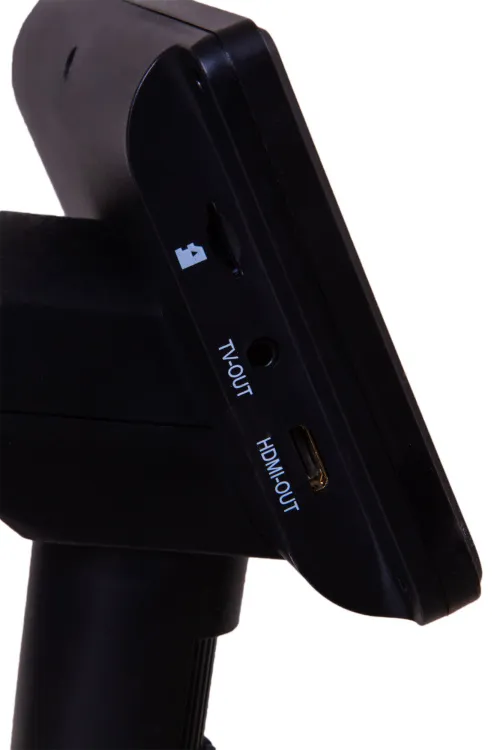
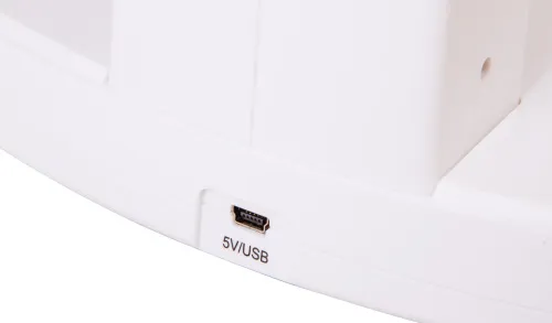
This self-sufficient microscope does not require connecting to external devices. A memory card is required for saving photos and videos (purchased separately). The microscope can be connected to an external monitor, a TV, or a computer. The kit includes all the necessary cables and software. Note: the software that is installed in a computer allows for measuring objects (line, radius, diameter, three-point angle). This software also allows for adding extra images and texts on top of images captured by a microscope.
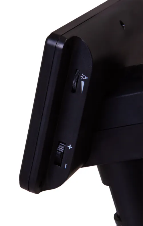
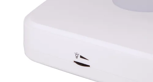
The illumination system of the microscope consists of bright LED lights with adjustable brightness. There is a built-in polarizing filter, which reduces parasitic reflections when studying shiny metals.
The microscope is powered by an AC supply or a built-in battery, which provides up to three hours of continuous work.
Features:
- Digital microscope with a 5M camera and a color LCD screen
- LED illumination with brightness adjustment
- Built-in polarizing filter
- Ability to record images and videos; an image is transmitted to an external screen
- Image processing software with measurement function
- Powered by an AC power supply or the built-in battery
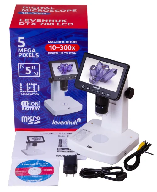
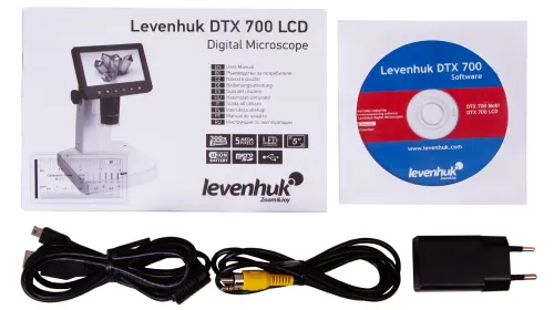
The kit includes:
- Microscope
- Adapter (100–240 V, 50/60 Hz)
- USB cable
- AV cable
- Calibration scale
- Software CD
- User manual and lifetime warranty
Recommendations on using the software: in order for the PortableCapture Plus program (and its other versions) to operate correctly, please launch the installed application only when your microscope is connected to a PC and ready for observations.
Caution! Remember that mains voltage in the U.S. and Canada is 110V and 220-240V in most European countries. Please refer to the specifications table for the correct mains voltage and never attempt to plug a 110V device into 220V outlet and vice versa without using a converter.
Levenhuk DTX 700 LCD Microscope is not compatible with external digital cameras.
| Product ID | 75075 |
| Brand | Levenhuk, Inc., USA |
| Warranty | lifetime |
| EAN | 5905555004389 |
| Package size (LxWxH) | 31x20x19 cm |
| Shipping Weight | 1.52 kg |
| Type | digital |
| Microscope head type | digital screen/PC monitor |
| Optics material | optical glass |
| Head | 5'' color LCD screen (fixed) |
| Magnification, x | 10–300 (optical), 10–1200 (digital) |
| Stage, mm | ø45 |
| Stage moving range, mm | fixed |
| Stage features | with clips |
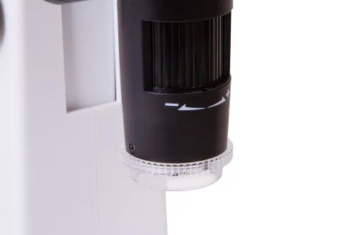
| Focus | manual, 5–70mm |
| Body | plastic |
| Illumination | LED |
| Brightness adjustment | ✓ |
| Power supply | 100–240V, 50/60Hz, 5V, 2A output, built-in 3.7V/2500mAh Li-ion battery |
| Power supply: batteries/built-in battery | yes |
| Light filters | polarizing |
| Operating temperature range, °C | -10...+65 |
| Additional | battery-operated work: 3 hours; charging time: 7 hours |
| Ability to connect additional equipment | microSD card up to 32 GB (not included) computer: via an USB cable (included) TV: via an AV cable (included) |
| User level | beginners, experienced users |
| Assembly and installation difficulty level | easy |
| Software language | Chinese, English, French, German, Japanese, Russian, Spanish |
| Application | for applied research |
| Illumination location | dual |
| Research method | bright field |
| Maximum resolution | images: 12Mpx, 10Mpx, 8Mpx, 5Mpx, 3Mpx, 2Mpx video: 1080p, 720p |
| Megapixels | 5 |
| Video recording | yes |
| Image format | *.jpg |
| Video format | *.avi |
| Frame rate | 30 frames per second |
| Software, drivers | photo and video capture and processing software with measurement function |
| Output | HDMI/TV, USB 2.0 |
| System requirements | OS: Windows 7/8/10, Mac 10.14 and later, minimum P2 1GHz, 512MB RAM, 512MB video card, USB 2.0 port, CD-ROM |
We have gathered answers to the most frequently asked questions to help you sort things out
Find out why studying eyes under a microscope is entertaining; how insects’ and arachnids’ eyes differ and what the best way is to observe such an interesting specimen
Read this review to learn how to observe human hair, what different hair looks like under a microscope and what magnification is required for observations
Learn what a numerical aperture is and how to choose a suitable objective lens for your microscope here
Learn what a spider looks like under microscope, when the best time is to take photos of it, how to study it properly at magnification and more interesting facts about observing insects and arachnids
This review for beginner explorers of the micro world introduces you to the optical, illuminating and mechanical parts of a microscope and their functions
Short article about Paramecium caudatum - a microorganism that is interesting to observe through any microscope
Best regards
Kaare Møller
thanks for your query.
You should choose 5M (default resolution) on the microscope.
Bring the stage near to the specimen and turn the fine focus wheel to the biggest magnification, then take a picture and save it to SD card. After that, import this picture into the PC software, and go to measurement function. Here the real size of object can be input to see the 300x magnification.
Hope this helps!

