BG
BG  BY
BY  CY
CY  CZ
CZ  DE
DE  EE
EE  ES
ES  GR
GR  HU
HU  IS
IS  IT
IT  LT
LT  LV
LV  MY
MY  PL
PL  PT
PT  RO
RO  SK
SK  TR
TR  UA
UA  USA
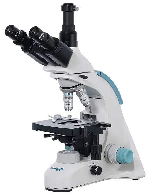
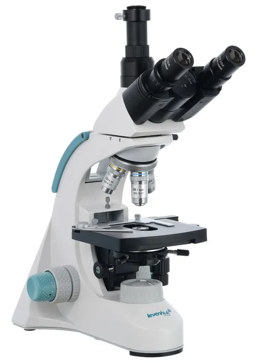
USA Levenhuk D900T Digital Trinocular Microscope
Magnification: 40–1000x. Trinocular head, achromatic objectives, and LED illumination with a collector lens
| Product ID | 75437 |
| Brand | Levenhuk, Inc., USA |
| Warranty | lifetime |
| EAN | 5905555005362 |
| Package size (LxWxH) | 51x25x40 cm |
| Shipping Weight | 6.64 kg |
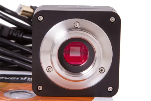
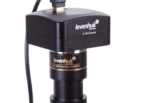
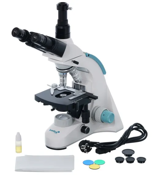
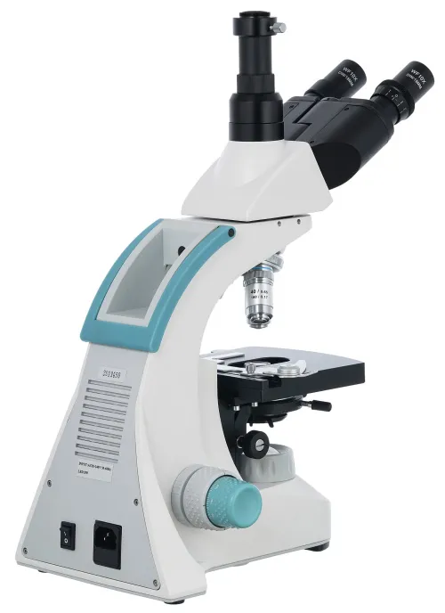
Levenhuk D900T is a trinocular microscope with a 5.1MP digital camera. It can be utilized for the laboratory research of any level of complexity as well as capturing photos and videos. The digital camera allows for taking high-resolution photos and videos of the observed object. It also transmits the image to the screen online. The image resolution reaches 2592х1944 pixels. This microscope is a great instrument for demanding professionals in different fields of science.
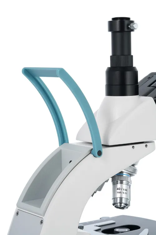
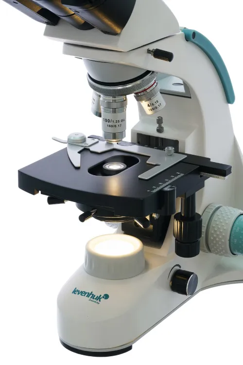
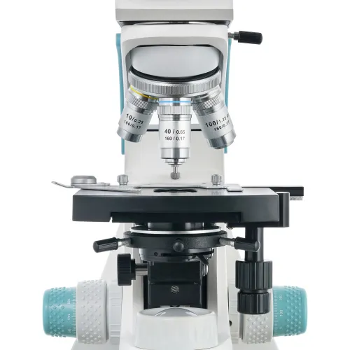
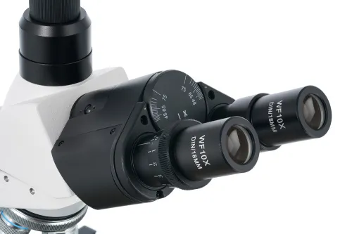
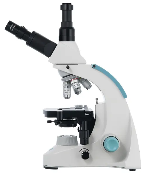
The optical scheme includes wide-field eyepieces with 10x magnification and achromatic objectives with various magnifications. The front lenses of the 40x and 100x objectives are equipped with protective spring-loaded frames. You can use the microscope for classic "dry" observations (all of the objectives) as well as observations using oil immersion (only 100x objective). The trinocular head can be rotated by 360°, and the visual module is slightly inclined by 30°. The digital camera (included) is installed in the third tube of the trinocular head.
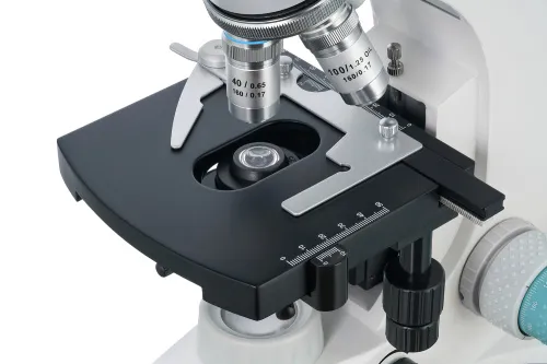
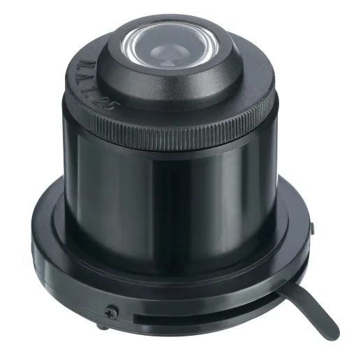
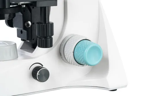
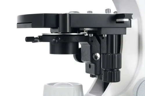
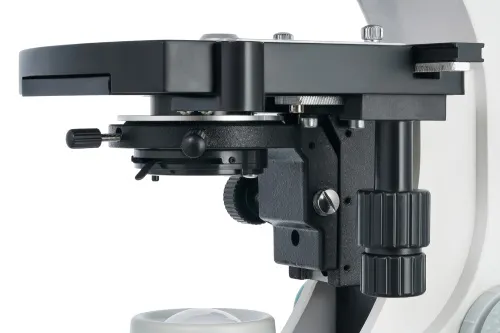
Samples for research are placed on a stage with a mechanical scale. The lighting system is located at the bottom and consists of the 3W LED and collector lens with adjustable brightness. The light intensity can also be adjusted using the Abbe condenser with an iris diaphragm and filter holder. The lighting system is powered by an AC power supply.
The digital camera is equipped with the necessary cables and software that enable you to crop the picture, resize it, adjust the contrast and brightness, and otherwise process images.
Note: Remember that the power supply in the US and Canada is 110V, and it is 220–240V in most European countries. Please refer to the specifications table for the correct voltage and never attempt to plug a 110V device into 220V outlet and vice versa without using a converter.
Some things that you can see under a microscope:


 under a microscope")
You can find these and many more interesting microscope slides to observe in the Levenhuk Prepared Slides Sets{:target="_blank"}.
The Levenhuk D900T Digital Trinocular Microscope is compatible with Levenhuk digital cameras{:target="_blank"} (additional cameras are purchased separately). Levenhuk cameras are installed in an eyepiece tube instead of an eyepiece.
Main Features:
- Wide-field achromatic optics
- Magnification: 40–1000x
- Lower 3 W LED lighting for observations in transmitted light
- Digital camera for photo and video recording of observations is included
- AC power supply
The kit includes:
- Microscope
- Achromatic objective lenses: 4x, 10x, 40x, and 100x (oil immersion)
- WF10x/18mm eyepieces (2 pcs)
- Abbe condenser N.A. 1.25 with an iris diaphragm and filter holder
- Filters: blue, green, yellow
- Fuse (2 pcs)
- C-mount
- Vial of immersion oil
- Power cord
- Dust cover
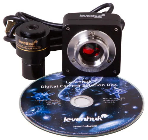
- Built-in 5.1MP digital camera
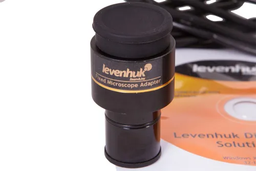
- Camera adapter
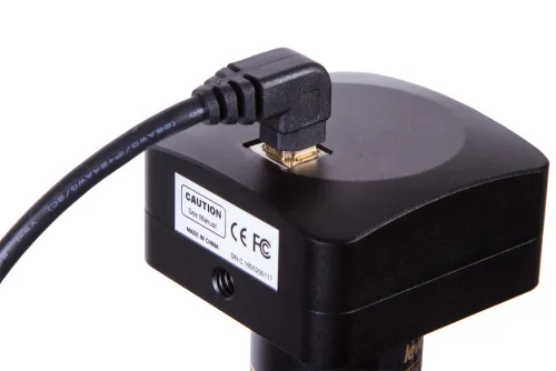
- USB cable
- Software CD and drivers
- User manual and lifetime warranty
| Product ID | 75437 |
| Brand | Levenhuk, Inc., USA |
| Warranty | lifetime |
| EAN | 5905555005362 |
| Package size (LxWxH) | 51x25x40 cm |
| Shipping Weight | 6.64 kg |
| Type | biological, light/optical, digital |
| Microscope head type | trinocular |
| Optics material | optical glass |
| Head | 360 ° rotatable |
| Head inclination angle | 30 ° |
| Magnification, x | 40 — 1000 |
| Eyepiece tube diameter, mm | 23.2 |
| Eyepieces | WF10x/18mm (2 pcs.) |
| Objectives | achromatic: 4x, 10x, 40xs, 100xs (oil immersion) |
| Revolving nosepiece | for 4 objectives |
| Interpupillary distance, mm | 48 — 75 |
| Stage, mm | 130x120 |
| Stage moving range, mm | 70/30 |
| Stage features | mechanical double-layer |
| Eyepiece diopter adjustment, diopters | ±5 |
| Condenser | Abbe N.A. 1.25 with an iris diaphragm and filter holder |
| Diaphragm | iris |
| Focus | coaxial, coarse (25mm) and fine (0.002mm) |
| Body | metal |
| Illumination | LED |
| Brightness adjustment | ✓ |
| Power supply | 110–220V |
| Light source type | with a collector, 3W LED |
| Light filters | blue, green, yellow |
| Application | laboratory/medical |
| Illumination location | lower |
| Research method | bright field |
| Digital camera included | ✓ |
| Maximum resolution | 2592x1944 |
| Camera specifications | |
| Megapixels | 5.1 |
| Pixel size, μm | 2.2x2.2 |
| Video recording | yes |
| Image format | *.bmp, *.jpeg, *.jpg, *.png, *.tif, *.tiff, *.gif, *.psd, *.ico, *.emf, *.wmf, etc. (photo); *.wmv, *.h264, *.avi etc. (video) |
| Spectral range, nm | 380–650 |
| White balance | automatic, manual |
| Exposure control | automatic, manual |
| Sensitivity, V/lux-sec@550nm | 0.53 |
| Usage location | eyepiece tube (replaces an eyepiece) |
| Method of exposure | ERS (Electronic Rolling Shutter) |
| Software, drivers | Levenhuk software, USB 2.0 driver |
| Programmable options | brightness, exposure control, image size |
| Output | 480 Mbps, USB 2.0 |
| System requirements | OS Windows XP/Vista/7/8/10/11 (32 and 64 bit), Mac 10.12, Linux Ubuntu 14.04; 2.8GHz Intel Core 2 and above, 2GB RAM, USB 2.0, CD-ROM |
| Camera power supply | via USB cable |
We have gathered answers to the most frequently asked questions to help you sort things out
Find out why studying eyes under a microscope is entertaining; how insects’ and arachnids’ eyes differ and what the best way is to observe such an interesting specimen
Read this review to learn how to observe human hair, what different hair looks like under a microscope and what magnification is required for observations
Learn what a numerical aperture is and how to choose a suitable objective lens for your microscope here
Learn what a spider looks like under microscope, when the best time is to take photos of it, how to study it properly at magnification and more interesting facts about observing insects and arachnids
This review for beginner explorers of the micro world introduces you to the optical, illuminating and mechanical parts of a microscope and their functions
Short article about Paramecium caudatum - a microorganism that is interesting to observe through any microscope