BG
BG  BY
BY  CY
CY  CZ
CZ  DE
DE  EE
EE  ES
ES  GR
GR  HU
HU  IS
IS  IT
IT  LT
LT  LV
LV  MY
MY  PL
PL  PT
PT  RO
RO  SK
SK  TR
TR  UA
UA  USA
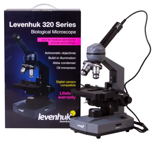
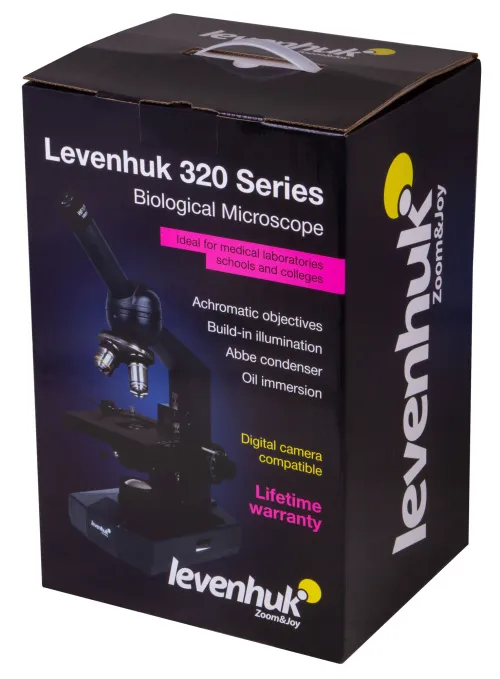
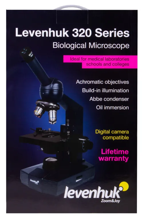
USA Levenhuk D320L BASE 3M Digital Monocular Microscope
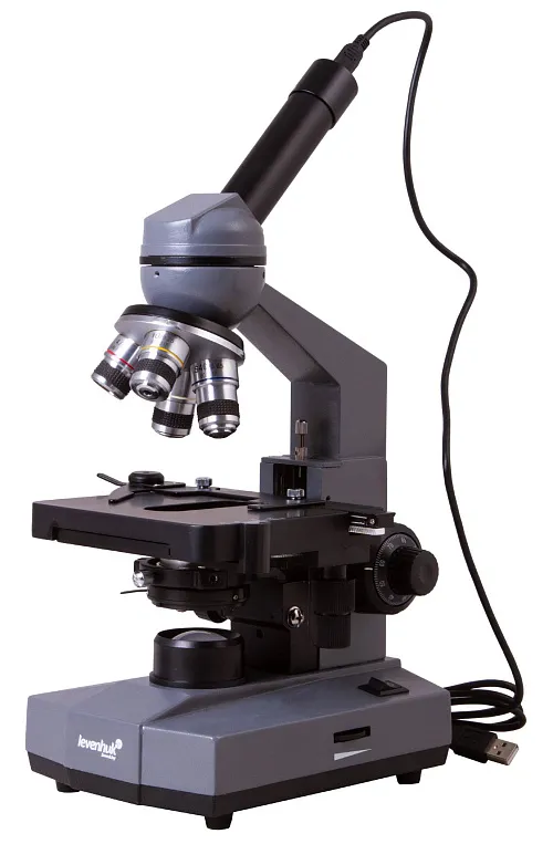


Monocular laboratory microscope with a digital camera and halogen illumination. Magnification: 40–1000x
| Product ID | 73812 |
| Brand | Levenhuk, Inc., USA |
| Warranty | lifetime |
| EAN | 5905555004525 |
| Package size (LxWxH) | 39x25x21 cm |
| Shipping Weight | 3.4 kg |
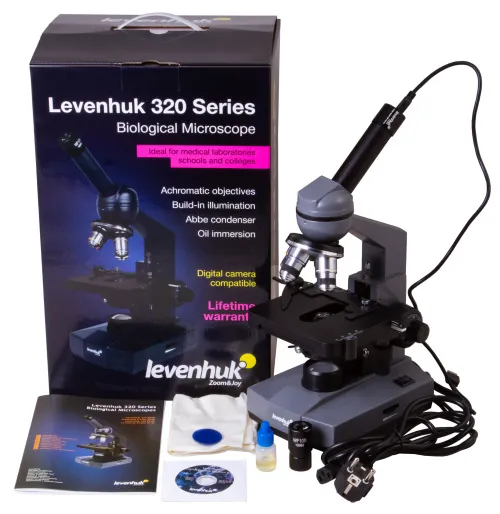
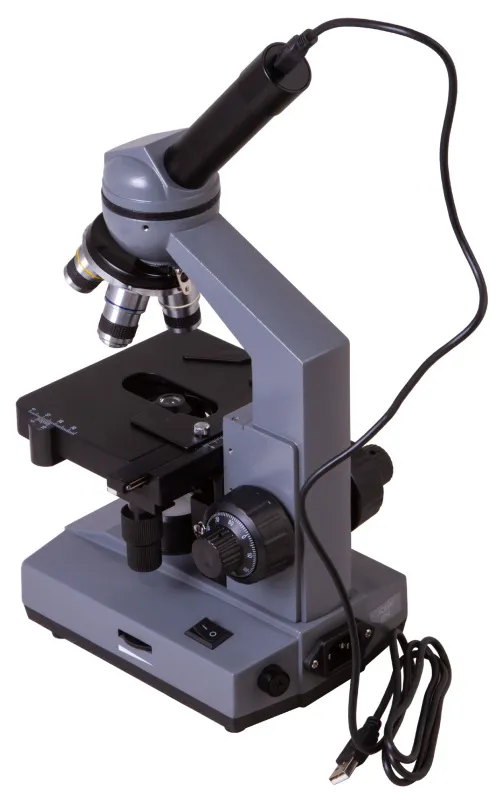
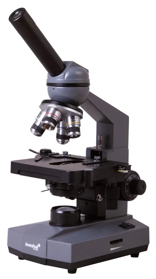
Levenhuk D320L BASE is a digital microscope with a 3M camera. It is great for performing observations in medical and clinical and diagnostic laboratories, holding seminars and lectures in academies and universities as well as in colleges, creating digital observational records. This microscope is very interesting to specialists of different scientific fields. It will become an excellent choice for professional microscopy observations.
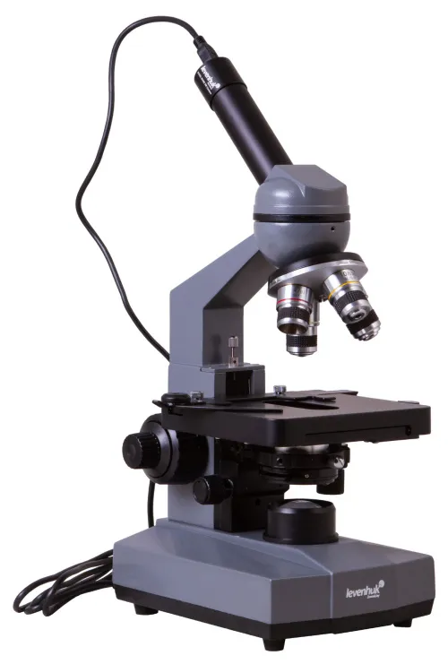
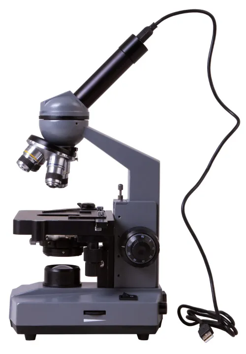
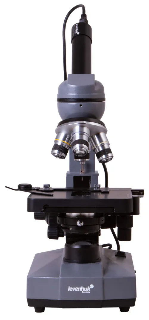
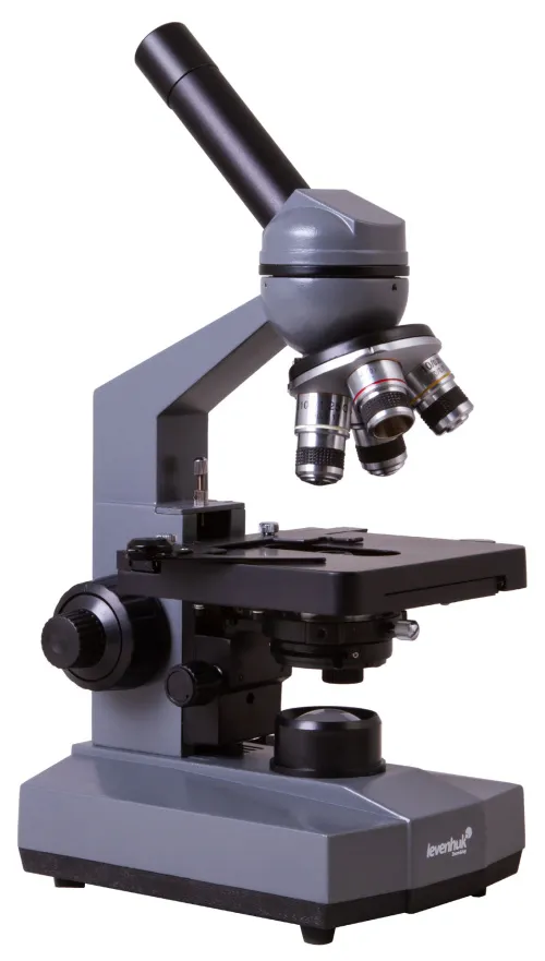
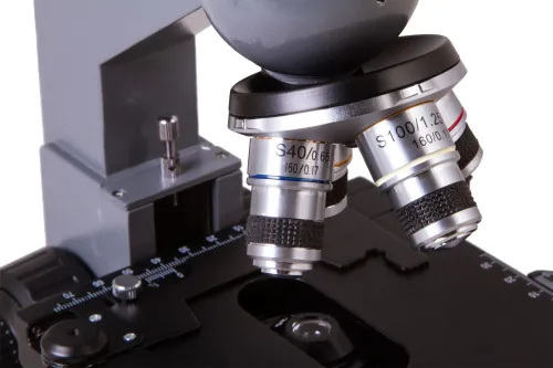
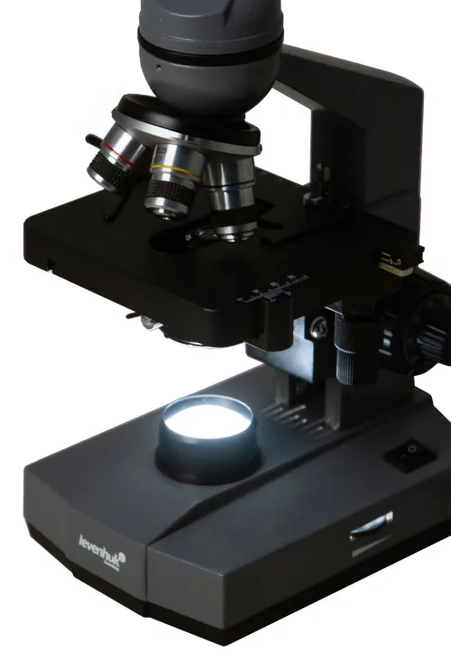
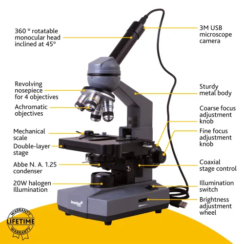
Levenhuk D320L BASE is a monocular microscope with a 360° rotatable head. A wide-angle 10x eyepiece and four achromatic objectives of different magnifications are used for observations. The magnification range varies from 40x to 1000x. You can apply oil immersion at maximum magnification (when using a 100x objective). A bottle of immersion oil comes included in the kit.
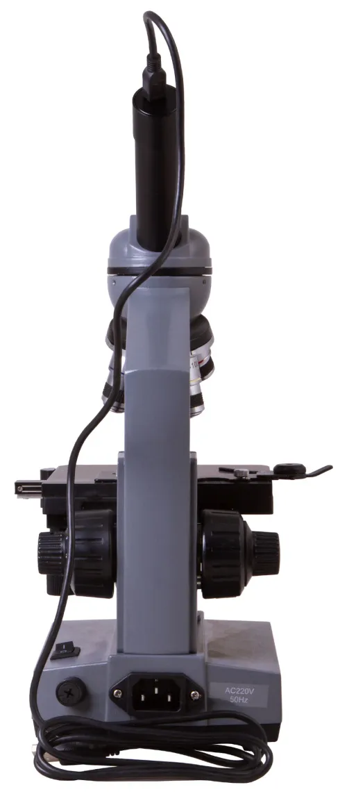
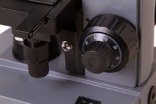
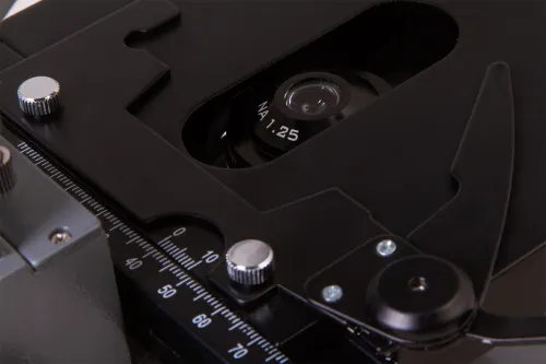
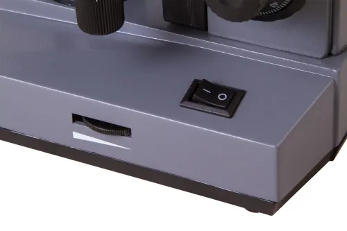
The illumination system is located in the lower part of the microscope. It consists of a powerful halogen lamp with adjustable brightness and an Abbe condenser with an iris diaphragm. Illumination is powered by an AC supply. You can use light filters for adjusting image contrast. Specimens are fixed on a stage, which is equipped with a mechanical scale. Sharpness adjustment is performed by rotating coarse and fine focusing adjustment knobs.
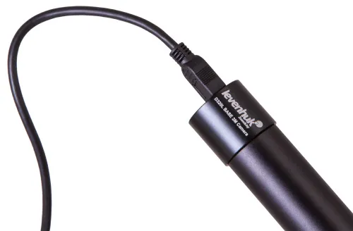
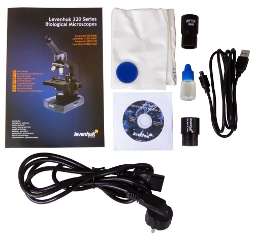
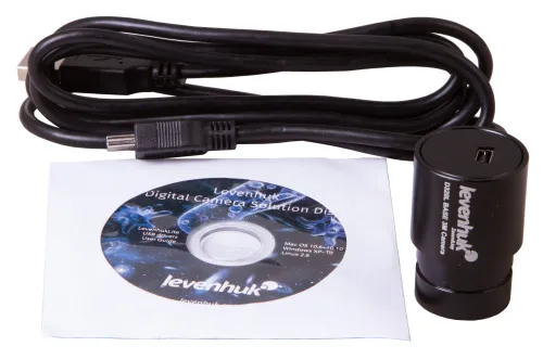
The kit includes a 3M digital camera. It allows for transmitting an image from a microscope to an external screen, e.g. a computer screen. You can make videos, take snapshots and process them with a special software program (an installation CD is included). The camera is powered by a computer with the help of a USB cable.
Features:
- Laboratory microscopes for observations in the transmitted light
- Rotatable monocular head, achromatic optics
- Magnification range: 40–1000x
- 3M digital camera is included in the kit
- 20W halogen illumination with brightness adjustment powered by an AC supply
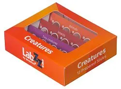
The kit includes:
- Microscope
- Achromatic objective lenses: 4x, 10x, 40x and 100x (oil immersion)
- Wide-angle WF10x eyepiece
- Blue filter
- Bottle of immersion oil
- AC adapter
- Dust cover
- Digital camera
- USB cable for camera
- Software CD
- User manual and lifetime warranty
Some things you can see under a microscope:


Levenhuk D320L BASE Microscope is compatible with Levenhuk digital cameras (purchased separately). Levenhuk cameras are installed in the eyepiece tube instead of an eyepiece.
| Product ID | 73812 |
| Brand | Levenhuk, Inc., USA |
| Warranty | lifetime |
| EAN | 5905555004525 |
| Package size (LxWxH) | 39x25x21 cm |
| Shipping Weight | 3.4 kg |
| Type | biological, light/optical, digital |
| Microscope head type | monocular |
| Optics material | optical glass |
| Head | 360 ° rotatable |
| Head inclination angle | 45 ° |
| Magnification, x | 40 — 1000 |
| Eyepiece tube diameter, mm | 23.2 |
| Eyepieces | WF10x |
| Objectives | achromatic: 4x, 10x, 40xs, 100xs (oil immersion) |
| Revolving nosepiece | for 4 objectives |
| Stage, mm | 110x125 |
| Stage moving range, mm | 58/25 |
| Stage features | mechanical double-layer |
| Condenser | Abbe N. A. 1.25 with an iris diaphragm |
| Diaphragm | iris |
| Focus | coaxial, coarse (20mm) and fine (0.002mm) |
| Body | metal |
| Illumination | halogen |
| Brightness adjustment | ✓ |
| Power supply | 220V/50Hz |
| Light source type | 20W |
| Light filters | blue |
| Additional | immersion oil included |
| User level | experienced users |
| Assembly and installation difficulty level | easy |
| Software language | Mac and Linux: English, Windows: English, Russian, French, German, Polish, Chinese, Turkish |
| Application | laboratory/medical |
| Illumination location | lower |
| Research method | bright field |
| Digital camera included | ✓ |
| Pouch/case/bag in set | dust cover |
| Maximum resolution | 2048x1536 |
| Camera specifications | |
| Megapixels | 3 |
| Sensor element | 1/2.7" |
| Pixel size, μm | 2.2x2.2 |
| Video recording | yes |
| Image format | *.jpg, *.bmp, *.png, *.tif and others |
| Video format | output: *.wmv, *.avi, *.h264 (Windows 8 and later), *h265 (Windows 10 and later) |
| Spectral range, nm | 380–650 |
| White balance | automatic, manual |
| Exposure control | automatic |
| Frame rate | 3@2048x1536 5@1600x1200 7.5@1280x1024 |
| Usage location | ocular tube, instead of the eyepiece |
| Method of exposure | ERS (Electronic Rolling Shutter) |
| Software, drivers | LevenhukLite |
| Programmable options | brightness, image size |
| Output | USB 2.0 |
| System requirements | Windows 8/10/11 (32bit and 64bit), Mac OS X, Linux, up to 2.8GHz Intel Core 2 or higher, minimum 2GB RAM, USB 2.0 port, CD-ROM |
| Camera power supply | via USB cable |
We have gathered answers to the most frequently asked questions to help you sort things out
Find out why studying eyes under a microscope is entertaining; how insects’ and arachnids’ eyes differ and what the best way is to observe such an interesting specimen
Read this review to learn how to observe human hair, what different hair looks like under a microscope and what magnification is required for observations
Learn what a numerical aperture is and how to choose a suitable objective lens for your microscope here
Learn what a spider looks like under microscope, when the best time is to take photos of it, how to study it properly at magnification and more interesting facts about observing insects and arachnids
This review for beginner explorers of the micro world introduces you to the optical, illuminating and mechanical parts of a microscope and their functions
Short article about Paramecium caudatum - a microorganism that is interesting to observe through any microscope