BG
BG  BY
BY  CY
CY  CZ
CZ  DE
DE  EE
EE  ES
ES  GR
GR  HU
HU  IS
IS  IT
IT  LT
LT  LV
LV  MY
MY  PL
PL  PT
PT  RO
RO  SK
SK  TR
TR  UA
UA  USA
USA Bresser Researcher LCD Microscope
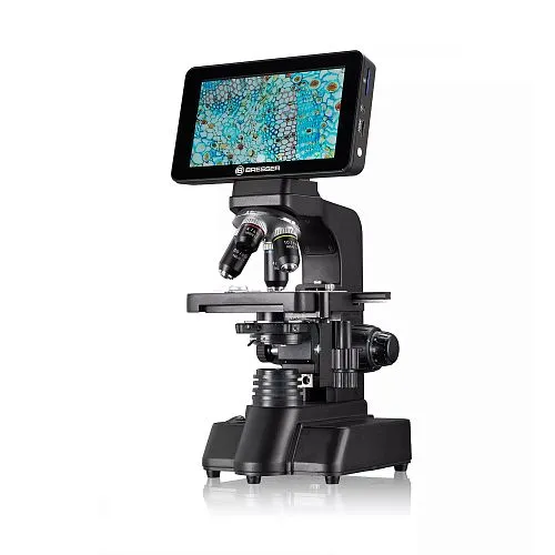
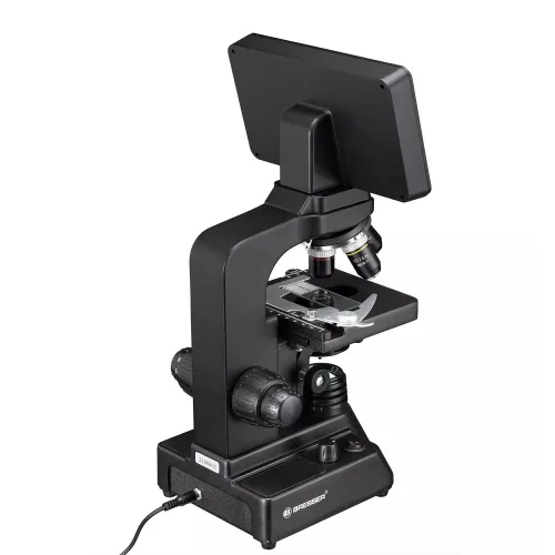
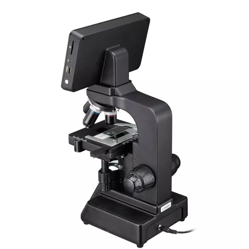
Digital microscope. Magnification: 40–600x
| Product ID | 78981 |
| Brand | Bresser GmbH, Germany |
| Warranty | 2 years |
| EAN | 4007922074887 |
| Package size (LxWxH) | 26x29x46 cm |
| Shipping Weight | 4.05 kg |
A digital microscope easily brings the microcosmos up on the screen. For a long time, these instruments were common only in the beginner's range. Now, the successful and proven Researcher series makes the step into the digital microscopy world and offers new options for microscopy in schools, for hobby or work. The optics and mechanics is the same as already well estabished in the Researcher Bino and Researcher Trino microscopes. Incuding 4 DIN objectives, Abbe condenser, coaxial fine focus and crosstable, the Researcher LCD is suitable for use in the microscopy hobby as well as in teaching or other work related applications. The highest magnification when using the 60x objective (compares to 600x on non digital microscopes) is high enough to view small microorganisms as microalgae, flagellates and bacteria. Other magnifications (20x, 100x oil) can be added later. The Researcher LCD is also compatible with our plan-achromatic objectives designed for 160mm optical tube length.
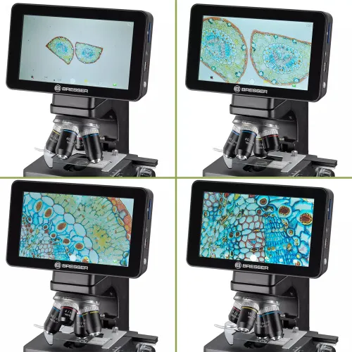
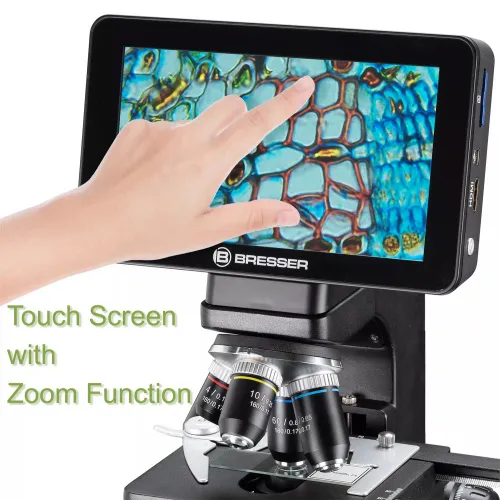
The integrated, modern Sony camerasensor IMX206 with 16MP gives a brilliant image with good contrast on the monitor. The user can change settings and capture images and video conveniently with the touch screen. Images with 16MP and Full HD video can be stored directly on SD card (SD card not supplied). The microscope also has a HDMI connection, so the image is easily shown on a big monitor, TV, beamer or smartboard. The results can be shared with a bigger audience in lectures or teaching.
Key features:
- Digital microscope with integrated 7'' touchscreen
- 4 achromatic DIN objectives (RMS thread connection), parfocal length 45mm
- Objective magnifications of 4x, 10x, 40x, 60x (40x and 60x with specimen protection)
- Abbe condenser with iris diaphragm, fully adjustable and height adjustable
- High-contrast screen with natural colours, 1024x600px resolution
- Integrated Sony IMX206 camera sensor with 16MP
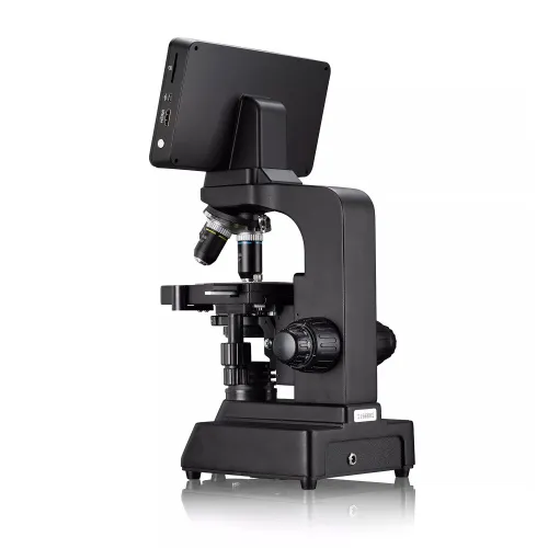
- HDMI port for connection to an external monitor
- SD card slot (supports up to 32GB, not included)
- USB port (for data transfer to SD card)
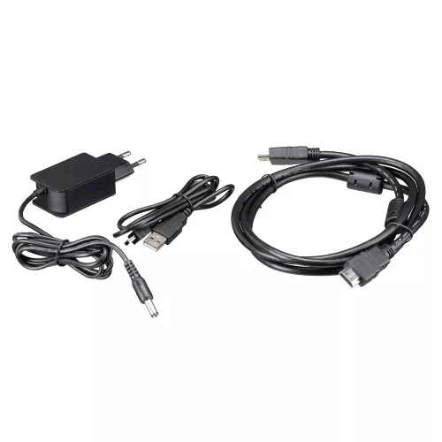
The kit includes:
- Researcher LCD digital microscope
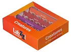
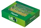
- Set of 5 prepared slides and 5 blank slides
- Power adapter (EU) 5V 2A
- HDMI cable
- USB cable for transferring stored data to a PC
- Dust cover
| Product ID | 78981 |
| Brand | Bresser GmbH, Germany |
| Warranty | 2 years |
| EAN | 4007922074887 |
| Package size (LxWxH) | 26x29x46 cm |
| Shipping Weight | 4.05 kg |
| Type | biological, light/optical |
| Microscope head type | binocular |
| Magnification, x | 40 — 600 |
| Objectives | 4x, 10x, 40x, 60x (achromat DIN standard) |
| Condenser | Abbe |
| Diaphragm | iris |
| Focus | coarse and fine |
| Body | aluminum |
| Illumination | LED |
| Brightness adjustment | ✓ |
| Power supply | via power adapter 5V, 2A |
| Application | school/educational, laboratory/medical |
| Pouch/case/bag in set | dust cover |
and downloads
We have gathered answers to the most frequently asked questions to help you sort things out
Find out why studying eyes under a microscope is entertaining; how insects’ and arachnids’ eyes differ and what the best way is to observe such an interesting specimen
Read this review to learn how to observe human hair, what different hair looks like under a microscope and what magnification is required for observations
Learn what a numerical aperture is and how to choose a suitable objective lens for your microscope here
Learn what a spider looks like under microscope, when the best time is to take photos of it, how to study it properly at magnification and more interesting facts about observing insects and arachnids
This review for beginner explorers of the micro world introduces you to the optical, illuminating and mechanical parts of a microscope and their functions
Short article about Paramecium caudatum - a microorganism that is interesting to observe through any microscope