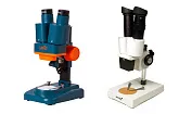
Microscope Slides Preparation Technique
Microscopes allow us to peek into the tiniest worlds. With a microscope you can open up a whole new world that exists at the cellular level: amazing, unpredictable and full of surprises.
One of the great things about microscope slides is that they can be stored for a long time and you can observe and study the process of cell decay over time. But if you can’t wait to observe some new interesting samples or if you are planning on major microscopic research, you will need to learn how to prepare microscope slides yourself.
Preparing dry mount microscope slides
Dry mount microscope slides are used for studying samples that don’t require contact with water in order to survive. The first thing you need to have is a clean blank slide. Very carefully place as thin as possible slice of a sample in the center of the blank slide and cover it with a cover glass. If you wear rubber gloves, you can gently pin down the cover glass to align the sample.
Preparing wet mount microscope slides
Wet mount microscope slides are used for studying samples, which can’t survive without water. Usually those are unicellular organisms and tiny animals. Take a clean blank slide. Place one or two drops of distilled water in the center of the slide using a dropper. Put the sample in the water and cover it with a cover glass. You can pin down the cover glass a little if you are wearing rubber gloves. Don’t touch the glass if you are not wearing gloves as fingerprints will certainly appear on the glass, which will significantly impede studying the sample.
A wet mount microscope slide holds the cover glass in place by itself and may be stored for some time. If observed microorganisms are too mobile to be properly studied, you can “slow them down” by adding a binder in the water, for example ProtoSlo (methyl cellulose in a 1.5% solution).
Staining samples
Some organisms are difficult to observe under a microscope without additional staining. One of the best ways to stain microscope a slide is to add a drop of Lugol's iodine (a water solution of iodine and potassium iodide) in the water before placing the sample in it. You can also use solutions of methylene blue or Gram crystal violet.
If you need to stain a prepared microscope slide, try the following trick. Place a drop of stain on one side of the cover glass and a small piece of napkin or a paper towel on the other side right up against the edge of the cover glass. The paper will draw the water from under the glass on one side and will also draw the stain under the cover glass on the other side.
Any reproduction of the material for public publication in any information medium and in any format is prohibited. You can refer to this article with active link to eu.levenhuk.com.
The manufacturer reserves the right to make changes to the pricing, product range and specifications or discontinue products without prior notice.


